


The crystal structure of thymidylate kinase from Pseudomonas aeruginosa PAO1 in complex with AZT Monophosphate
Tan, K., Joachimiak, G., Jedrzejczak, R., Sacchettini, J., Joachimiak, A.To be published.
Experimental Data Snapshot
Starting Model: experimental
View more details
Entity ID: 1 | |||||
|---|---|---|---|---|---|
| Molecule | Chains | Sequence Length | Organism | Details | Image |
| Thymidylate kinase | 213 | Pseudomonas aeruginosa PAO1 | Mutation(s): 0 Gene Names: PA2962, tmk EC: 2.7.4.9 | 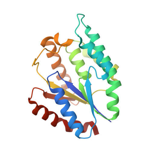 | |
UniProt | |||||
Entity Groups | |||||
| Sequence Clusters | 30% Identity50% Identity70% Identity90% Identity95% Identity100% Identity | ||||
| UniProt Group | Q9HZN8 | ||||
Sequence AnnotationsExpand | |||||
Reference Sequence | |||||
| Ligands 4 Unique | |||||
|---|---|---|---|---|---|
| ID | Chains | Name / Formula / InChI Key | 2D Diagram | 3D Interactions | |
| ATM Download:Ideal Coordinates CCD File | E [auth A], H [auth B], L [auth C], O [auth D] | 3'-AZIDO-3'-DEOXYTHYMIDINE-5'-MONOPHOSPHATE C10 H14 N5 O7 P OIFWQOKDSPDILA-XLPZGREQSA-N |  | ||
| GOL Download:Ideal Coordinates CCD File | G [auth A], J [auth B], K [auth B] | GLYCEROL C3 H8 O3 PEDCQBHIVMGVHV-UHFFFAOYSA-N |  | ||
| CA Download:Ideal Coordinates CCD File | P [auth D] | CALCIUM ION Ca BHPQYMZQTOCNFJ-UHFFFAOYSA-N |  | ||
| CL Download:Ideal Coordinates CCD File | F [auth A] I [auth B] M [auth C] N [auth C] Q [auth D] | CHLORIDE ION Cl VEXZGXHMUGYJMC-UHFFFAOYSA-M |  | ||
| Length ( Å ) | Angle ( ˚ ) |
|---|---|
| a = 44.5 | α = 90 |
| b = 123.404 | β = 100 |
| c = 73.92 | γ = 90 |
| Software Name | Purpose |
|---|---|
| SBC-Collect | data collection |
| MOLREP | phasing |
| PHENIX | refinement |
| HKL-3000 | data reduction |
| HKL-3000 | data scaling |


