A study on the interaction of rhodamine B with methylthioadenosine phosphorylase protein sourced from an antarctic soil metagenomic library.
Bartasun, P., Cieslinski, H., Bujacz, A., Wierzbicka-Wos, A., Kur, J.(2013) PLoS One 8: e55697-e55697
- PubMed: 23383268 Search on PubMedSearch on PubMed Central
- DOI: https://doi.org/10.1371/journal.pone.0055697
- Primary Citation Related Structures:
4GLF, 4GLJ - PubMed Abstract:
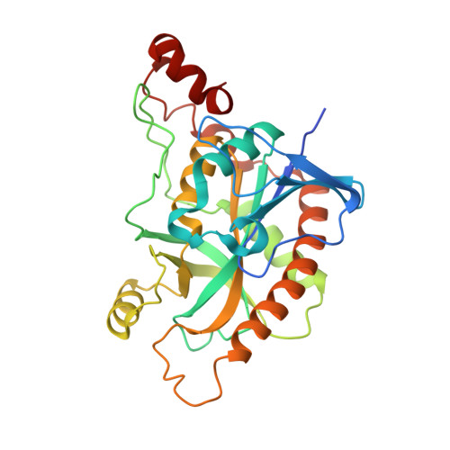
The presented study examines the phenomenon of the fluorescence under UV light excitation (312 nm) of E. coli cells expressing a novel metagenomic-derived putative methylthioadenosine phosphorylase gene, called rsfp, grown on LB agar supplemented with a fluorescent dye rhodamine B. For this purpose, an rsfp gene was cloned and expressed in an LMG194 E. coli strain using an arabinose promoter. The resulting RSFP protein was purified and its UV-VIS absorbance spectrum and emission spectrum were assayed. Simultaneously, the same spectroscopic studies were carried out for rhodamine B in the absence or presence of RSFP protein or native E. coli proteins, respectively. The results of the spectroscopic studies suggested that the fluorescence of E. coli cells expressing rsfp gene under UV illumination is due to the interaction of rhodamine B molecules with the RSFP protein. Finally, this interaction was proved by a crystallographic study and then by site-directed mutagenesis of rsfp gene sequence. The crystal structures of RSFP apo form (1.98 Å) and complex RSFP/RB (1.90 Å) show a trimer of RSFP molecules located on the crystallographic six fold screw axis. The RSFP complex with rhodamine B revealed the binding site for RB, in the pocket located on the interface between symmetry related monomers.
- Department of Microbiology, Faculty of Chemistry, Gdańsk University of Technology, Gdańsk, Poland.
Organizational Affiliation: