4GK5

Crystal structure of human Rev3-Rev7-Rev1-Polkappa complex


Experimental Data Snapshot

  • Method: X-RAY DIFFRACTION
  • Resolution: 3.21 Å
  • R-Value Free: 0.281 
  • R-Value Work: 0.232 
  • R-Value Observed: 0.234 

Starting Model: experimental
View more details

wwPDB Validation   3D Report Full Report


This is version 1.1 of the entry. See complete history


Literature

Structural insights into the assembly of human translesion polymerase complexes

Xie, W.Yang, X.Xu, M.Jiang, T.

(2012) Protein Cell 3: 864-874

  • DOI: https://doi.org/10.1007/s13238-012-2102-x
  • Primary Citation of Related Structures:  
    4GK0, 4GK5

  • PubMed Abstract: 

    In addition to DNA repair pathways, cells utilize translesion DNA synthesis (TLS) to bypass DNA lesions during replication. During TLS, Y-family DNA polymerase (Polη, Polκ, Polı and Rev1) inserts specific nucleotide opposite preferred DNA lesions, and then Polζ consisting of two subunits, Rev3 and Rev7, carries out primer extension. Here, we report the complex structures of Rev3-Rev7-Rev1(CTD) and Rev3-Rev7-Rev1(CTD)-Polκ(RIR). These two structures demonstrate that Rev1(CTD) contains separate binding sites for Polκ and Rev7. Our BIAcore experiments provide additional support for the notion that the interaction between Rev3 and Rev7 increases the affinity of Rev7 and Rev1. We also verified through FRET experiment that Rev1, Rev3, Rev7 and Polκ form a stable quaternary complex in vivo, thereby suggesting an efficient switching mechanism where the "inserter" polymerase can be immediately replaced by an "extender" polymerase within the same quaternary complex.


  • Organizational Affiliation

    National Laboratory of Biomacromolecules, Institute of Biophysics, Chinese Academy of Sciences, Chaoyang District, Beijing, China.


Macromolecules
Find similar proteins by:  (by identity cutoff)  |  3D Structure
Entity ID: 1
MoleculeChains Sequence LengthOrganismDetailsImage
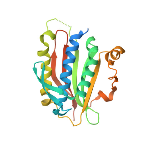
Mitotic spindle assembly checkpoint protein MAD2B
A, B
238Homo sapiensMutation(s): 1 
Gene Names: MAD2L2MAD2BREV7
UniProt & NIH Common Fund Data Resources
Find proteins for Q9UI95 (Homo sapiens)
Explore Q9UI95 
Go to UniProtKB:  Q9UI95
PHAROS:  Q9UI95
GTEx:  ENSG00000116670 
Entity Groups  
Sequence Clusters30% Identity50% Identity70% Identity90% Identity95% Identity100% Identity
UniProt GroupQ9UI95
Sequence Annotations
Expand
  • Reference Sequence
Find similar proteins by:  (by identity cutoff)  |  3D Structure
Entity ID: 2
MoleculeChains Sequence LengthOrganismDetailsImage
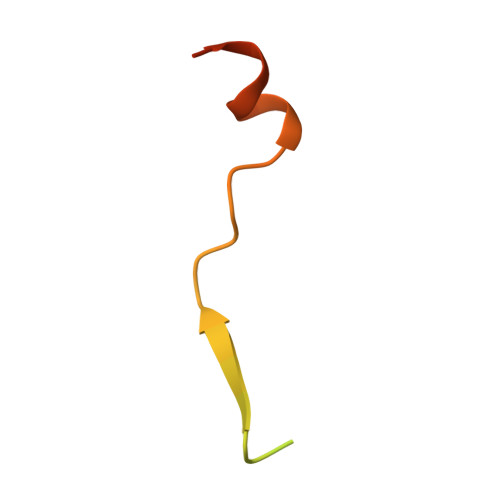
DNA polymerase zeta catalytic subunit
C, D
52Homo sapiensMutation(s): 0 
Gene Names: REV3LPOLZREV3
EC: 2.7.7.7
UniProt & NIH Common Fund Data Resources
Find proteins for O60673 (Homo sapiens)
Explore O60673 
Go to UniProtKB:  O60673
PHAROS:  O60673
GTEx:  ENSG00000009413 
Entity Groups  
Sequence Clusters30% Identity50% Identity70% Identity90% Identity95% Identity100% Identity
UniProt GroupO60673
Sequence Annotations
Expand
  • Reference Sequence
Find similar proteins by:  (by identity cutoff)  |  3D Structure
Entity ID: 3
MoleculeChains Sequence LengthOrganismDetailsImage
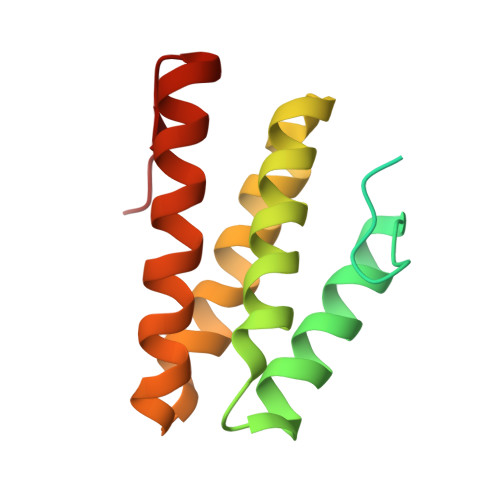
DNA repair protein REV1
E, F
136Homo sapiensMutation(s): 0 
Gene Names: REV1REV1L
EC: 2.7.7
UniProt & NIH Common Fund Data Resources
Find proteins for Q9UBZ9 (Homo sapiens)
Explore Q9UBZ9 
Go to UniProtKB:  Q9UBZ9
PHAROS:  Q9UBZ9
GTEx:  ENSG00000135945 
Entity Groups  
Sequence Clusters30% Identity50% Identity70% Identity90% Identity95% Identity100% Identity
UniProt GroupQ9UBZ9
Sequence Annotations
Expand
  • Reference Sequence

Find similar proteins by:  Sequence   |   3D Structure  

Entity ID: 4
MoleculeChains Sequence LengthOrganismDetailsImage
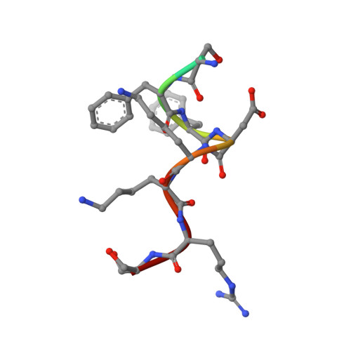
DNA polymerase kappa10Homo sapiensMutation(s): 0 
EC: 2.7.7.7
UniProt & NIH Common Fund Data Resources
Find proteins for Q9UBT6 (Homo sapiens)
Explore Q9UBT6 
Go to UniProtKB:  Q9UBT6
PHAROS:  Q9UBT6
GTEx:  ENSG00000122008 
Entity Groups  
Sequence Clusters30% Identity50% Identity70% Identity90% Identity95% Identity100% Identity
UniProt GroupQ9UBT6
Sequence Annotations
Expand
  • Reference Sequence
Experimental Data & Validation

Experimental Data

  • Method: X-RAY DIFFRACTION
  • Resolution: 3.21 Å
  • R-Value Free: 0.281 
  • R-Value Work: 0.232 
  • R-Value Observed: 0.234 
  • Space Group: P 21 21 2
Unit Cell:
Length ( Å )Angle ( ˚ )
a = 119.998α = 90
b = 72.762β = 90
c = 104.653γ = 90
Software Package:
Software NamePurpose
ADSCdata collection
MOLREPphasing
REFMACrefinement
HKL-2000data reduction
HKL-2000data scaling

Structure Validation

View Full Validation Report


Entry History 

Deposition Data

Revision History  (Full details and data files)

  • Version 1.0: 2013-03-13
    Type: Initial release
  • Version 1.1: 2023-11-08
    Changes: Data collection, Database references, Refinement description