4GIN | pdb_00004gin

Crystal structure of the MUTB R284C mutant from crystals soaked with the inhibitor deoxynojirimycin


Experimental Data Snapshot

  • Method: X-RAY DIFFRACTION
  • Resolution: 1.90 Å
  • R-Value Free: 
    0.181 (Depositor), 0.181 (DCC) 
  • R-Value Work: 
    0.140 (Depositor), 0.141 (DCC) 
  • R-Value Observed: 
    0.144 (Depositor) 

Starting Model: experimental
View more details

wwPDB Validation 3D Report Full Report

Validation slider image for 4GIN

This is version 1.2 of the entry. See complete history

Literature

Mutations inducing an active-site aperture in Rhizobium sp. sucrose isomerase confer hydrolytic activity

Lipski, A.Watzlawick, H.Ravaud, S.Robert, X.Rhimi, M.Haser, R.Mattes, R.Aghajari, N.

(2013) Acta Crystallogr D Biol Crystallogr 69: 298-307

  • DOI: https://doi.org/10.1107/S0907444912045532
  • Primary Citation Related Structures: 
    4GI6, 4GI8, 4GI9, 4GIA, 4GIN, 4H2C

  • PubMed Abstract: 

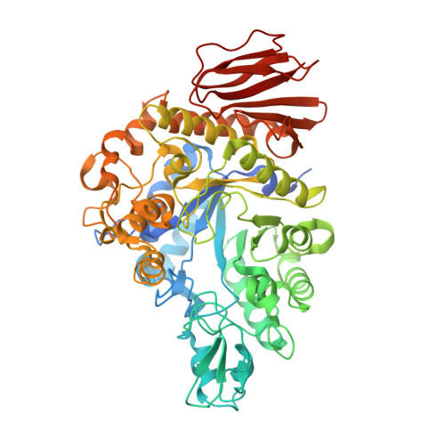
    Sucrose isomerase is an enzyme that catalyzes the production of sucrose isomers of high biotechnological and pharmaceutical interest. Owing to the complexity of the chemical synthesis of these isomers, isomaltulose and trehalulose, enzymatic conversion remains the preferred method for obtaining these products. Depending on the microbial source, the ratio of the sucrose-isomer products varies significantly. In studies aimed at understanding and explaining the underlying molecular mechanisms of these reactions, mutations obtained using a random-mutagenesis approach displayed a major hydrolytic activity. Two of these variants, R284C and F164L, of sucrose isomerase from Rhizobium sp. were therefore crystallized and their crystal structures were determined. The three-dimensional structures of these mutants allowed the identification of the molecular determinants that favour hydrolytic activity compared with transferase activity. Substantial conformational changes resulting in an active-site opening were observed, as were changes in the pattern of water molecules bordering the active-site region.


  • Organizational Affiliation
    • Laboratory for Biocrystallography and Structural Biology of Therapeutic Targets, Molecular and Structural Bases of Infectious Diseases, UMR 5086 CNRS and University of Lyon, 7 Passage du Vercors, F-69367 Lyon CEDEX 07, France.

Macromolecule Content 

  • Total Structure Weight: 67.22 kDa 
  • Atom Count: 5,246 
  • Modeled Residue Count: 555 
  • Deposited Residue Count: 584 
  • Unique protein chains: 1

Macromolecules

Find similar proteins by:|  3D Structure
Entity ID: 1
MoleculeChains  Sequence LengthOrganismDetailsImage
Sucrose isomerase584Rhizobium sp. MX-45Mutation(s): 1 
Gene Names: mutB
UniProt
Find proteins for M1E1F6 (Rhizobium sp. MX-45)
Explore M1E1F6 
Go to UniProtKB:  M1E1F6
Entity Groups
Sequence Clusters30% Identity50% Identity70% Identity90% Identity95% Identity100% Identity
UniProt GroupM1E1F6
Sequence Annotations
Expand
Reference Sequence

Experimental Data & Validation

Experimental Data

  • Method: X-RAY DIFFRACTION
  • Resolution: 1.90 Å
  • R-Value Free:  0.181 (Depositor), 0.181 (DCC) 
  • R-Value Work:  0.140 (Depositor), 0.141 (DCC) 
  • R-Value Observed: 0.144 (Depositor) 
Space Group: P 21 21 21
Unit Cell:
Length ( Å )Angle ( ˚ )
a = 83.8α = 90
b = 89.7β = 90
c = 91.6γ = 90
Software Package:
Software NamePurpose
ADSCdata collection
AMoREphasing
PHENIXrefinement
XDSdata reduction
XSCALEdata scaling

Structure Validation

View Full Validation Report


Entry History 

Deposition Data

Revision History  (Full details and data files)

  • Version 1.0: 2013-02-13
    Type: Initial release
  • Version 1.1: 2013-07-03
    Changes: Database references
  • Version 1.2: 2023-11-08
    Changes: Data collection, Database references, Derived calculations, Refinement description