Structural analysis of HmtT and HmtN involved in the tailoring steps of himastatin biosynthesis
Zhang, H., Chen, J., Wang, H., Xie, Y., Ju, J., Yan, Y., Zhang, H.(2013) FEBS Lett 587: 1675-1680
- PubMed: 23611984 Search on PubMed
- DOI: https://doi.org/10.1016/j.febslet.2013.04.013
- Primary Citation Related Structures:
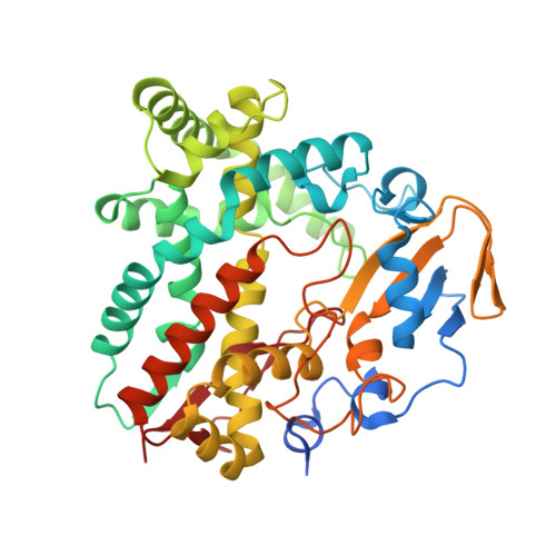
4GGV - PubMed Abstract:
Himastatin is a novel antibiotic featuring a bicyclohexadepsipeptide structure. On the himastatin biosynthesis pathway, three cytochrome P450s (HmtT, HmtN, HmtS) are responsible for the post-tailoring of the cyclohexadepsipeptide backbone. Here we report the crystal structures of HmtT and HmtN. The overall structures of these two proteins are homologous to other cytochrome P450s. However, the exceptionally long F-G loop in HmtT has a highly unusual conformation and extends deep into the active site. As a result, the F/G helices of HmtT are both kinked. In contrast, the F/G helices of HmtN are straight. Also, the F/G helices in HmtT and HmtN take distinctive orientations, which may be a contributing factor for the substrate specificity of these two enzymes.
- Key Laboratory of Molecular Biophysics, Ministry of Education, Wuhan 430074, Hubei, China.
Organizational Affiliation: