4GG7 | pdb_00004gg7

Crystal structure of cMET in complex with novel inhibitor


Experimental Data Snapshot

  • Method: X-RAY DIFFRACTION
  • Resolution: 2.27 Å
  • R-Value Free: 
    0.262 (Depositor), 0.266 (DCC) 
  • R-Value Work: 
    0.204 (Depositor), 0.207 (DCC) 
  • R-Value Observed: 
    0.210 (Depositor) 

wwPDB Validation 3D Report Full Report

Validation slider image for 4GG7

Ligand Structure Quality Assessment 


This is version 1.2 of the entry. See complete history

Literature

Multisubstituted quinoxalines and pyrido[2,3-d]pyrimidines: Synthesis and SAR study as tyrosine kinase c-Met inhibitors.

Wu, K.Ai, J.Liu, Q.Chen, T.Zhao, A.Peng, X.Wang, Y.Ji, Y.Yao, Q.Xu, Y.Geng, M.Zhang, A.

(2012) Bioorg Med Chem Lett 22: 6368-6372

  • DOI: https://doi.org/10.1016/j.bmcl.2012.08.075
  • Primary Citation Related Structures: 
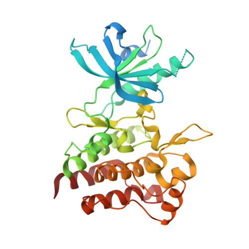
    4GG5, 4GG7

  • PubMed Abstract: 

    Two series of new analogues were designed by replacing the quinoline scaffold of our earlier lead 2 (zgw-atinib) with quinoxaline and pyrido[2,3-d]pyrimidine frameworks. Moderate c-Met inhibitory activity was observed in the quinoxaline series. Among the pyrido[2,3-d]pyrimidine series, compounds 13a-c possessing an O-linkage were inactive, whilst the N-linked analogues 15a-c retained c-Met inhibitory potency. Highest activity was observed in the 3-nitrobenzyl analog 15b that showed an IC(50) value of 6.5 nM. Further structural modifications based on this compound were undergoing.


  • Organizational Affiliation
    • Department of Medicinal Chemistry, China Pharmaceutical University, Nanjing 210009, China.

Macromolecule Content 

  • Total Structure Weight: 36.53 kDa 
  • Atom Count: 2,393 
  • Modeled Residue Count: 278 
  • Deposited Residue Count: 319 
  • Unique protein chains: 1

Macromolecules

Find similar proteins by:|  3D Structure
Entity ID: 1
MoleculeChains  Sequence LengthOrganismDetailsImage
Hepatocyte growth factor receptor319Homo sapiensMutation(s): 0 
Gene Names: CMETMET
EC: 2.7.10.1
UniProt & NIH Common Fund Data Resources
Find proteins for P08581 (Homo sapiens)
Explore P08581 
Go to UniProtKB:  P08581
PHAROS:  P08581
GTEx:  ENSG00000105976 
Entity Groups
Sequence Clusters30% Identity50% Identity70% Identity90% Identity95% Identity100% Identity
UniProt GroupP08581
Sequence Annotations
Expand
Reference Sequence

Small Molecules

Ligands 1 Unique
IDChains Name / Formula / InChI Key2D Diagram3D Interactions
0J8

Query on 0J8


Download:Ideal Coordinates CCD File
B [auth A]N-(3-nitrobenzyl)-6-[1-(piperidin-4-yl)-1H-pyrazol-4-yl]-2-(trifluoromethyl)pyrido[2,3-d]pyrimidin-4-amine
C23 H21 F3 N8 O2
OIYLUARXJZNDPY-UHFFFAOYSA-N

Experimental Data & Validation

Experimental Data

  • Method: X-RAY DIFFRACTION
  • Resolution: 2.27 Å
  • R-Value Free:  0.262 (Depositor), 0.266 (DCC) 
  • R-Value Work:  0.204 (Depositor), 0.207 (DCC) 
  • R-Value Observed: 0.210 (Depositor) 
Space Group: P 41 21 2
Unit Cell:
Length ( Å )Angle ( ˚ )
a = 65.867α = 90
b = 65.867β = 90
c = 187.176γ = 90
Software Package:
Software NamePurpose
XSCALEdata scaling
PHASERphasing
PHENIXrefinement
PDB_EXTRACTdata extraction
Blu-Icedata collection
XDSdata reduction

Structure Validation

View Full Validation Report


Ligand Structure Quality Assessment 


Entry History 

Deposition Data

Revision History  (Full details and data files)

  • Version 1.0: 2012-10-03
    Type: Initial release
  • Version 1.1: 2012-10-17
    Changes: Database references
  • Version 1.2: 2024-02-28
    Changes: Data collection, Database references, Derived calculations