4G9M | pdb_00004g9m

Crystal structure of the Rhizoctonia solani agglutinin


Experimental Data Snapshot

  • Method: X-RAY DIFFRACTION
  • Resolution: 1.60 Å
  • R-Value Free: 
    0.232 (Depositor), 0.228 (DCC) 
  • R-Value Work: 
    0.181 (Depositor), 0.177 (DCC) 
  • R-Value Observed: 
    0.184 (Depositor) 

Starting Model: experimental
View more details

wwPDB Validation 3D Report Full Report

Validation slider image for 4G9M

This is version 1.2 of the entry. See complete history

Literature

Structural analysis of the Rhizoctonia solani agglutinin reveals a domain-swapping dimeric assembly.

Skamnaki, V.T.Peumans, W.J.Kantsadi, A.L.Cubeta, M.A.Plas, K.Pakala, S.Zographos, S.E.Smagghe, G.Nierman, W.C.Van Damme, E.J.Leonidas, D.D.

(2013) FEBS J 280: 1750-1763

  • DOI: https://doi.org/10.1111/febs.12190
  • Primary Citation Related Structures: 
    4G9M, 4G9N

  • PubMed Abstract: 

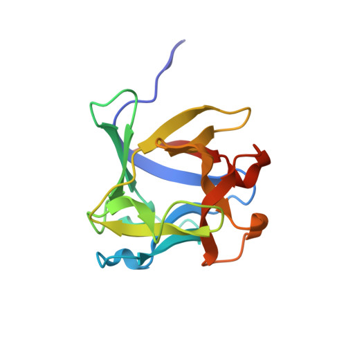
    Rhizoctonia solani agglutinin (RSA) is a 15.5-kDa lectin accumulated in the mycelium and sclerotia of the soil born plant pathogenic fungus R. solani. Although it is considered to serve as a storage protein and is implicated in fungal insecticidal activity, its physiological role remains unclear as a result of a lack of any structure/function relationship information. Glycan arrays showed that RSA displays high selectivity towards terminal nonreducing N-acetylgalactosamine residues. We determined the amino acid sequence of RSA and also determined the crystal structures of the free form and the RSA-N-acetylgalactosamine complex at 1.6 and 2.2 Å resolution, respectively. RSA is a homodimer comprised of two monomers adopting the β-trefoil fold. Each monomer accommodates two different carbohydrate-binding sites in an asymmetric way. Despite RSA topology similarities with R-type lectins, the two-monomer assembly involves an N-terminal swap, thus creating a dimer association novel to R-type lectins. Structural characterization of the two carbohydrate-binding sites offers insights on the structural determinants of the RSA carbohydrate specificity. Structural data have been deposited in the Protein Data Bank database under accession numbers 4G9M and 4G9N. RSA and RSA bind by x-ray crystallography (View interaction).


  • Organizational Affiliation
    • Department of Biochemistry and Biotechnology, University of Thessaly, Larissa, Greece.

Macromolecule Content 

  • Total Structure Weight: 30.97 kDa 
  • Atom Count: 2,443 
  • Modeled Residue Count: 285 
  • Deposited Residue Count: 286 
  • Unique protein chains: 1

Macromolecules

Find similar proteins by:|  3D Structure
Entity ID: 1
MoleculeChains  Sequence LengthOrganismDetailsImage
agglutinin
A, B
143Rhizoctonia solaniMutation(s): 0 
UniProt
Find proteins for L8WGI4 (Thanatephorus cucumeris (strain AG1-IA))
Explore L8WGI4 
Go to UniProtKB:  L8WGI4
Entity Groups
Sequence Clusters30% Identity50% Identity70% Identity90% Identity95% Identity100% Identity
UniProt GroupL8WGI4
Sequence Annotations
Expand
Reference Sequence

Experimental Data & Validation

Experimental Data

  • Method: X-RAY DIFFRACTION
  • Resolution: 1.60 Å
  • R-Value Free:  0.232 (Depositor), 0.228 (DCC) 
  • R-Value Work:  0.181 (Depositor), 0.177 (DCC) 
  • R-Value Observed: 0.184 (Depositor) 
Space Group: C 1 2 1
Unit Cell:
Length ( Å )Angle ( ˚ )
a = 121.979α = 90
b = 61.091β = 93
c = 32.793γ = 90
Software Package:
Software NamePurpose
DNAdata collection
MrBUMPphasing
PHENIXrefinement
DENZOdata reduction
SCALEPACKdata scaling

Structure Validation

View Full Validation Report


Entry History 

Deposition Data

Revision History  (Full details and data files)

  • Version 1.0: 2013-06-05
    Type: Initial release
  • Version 1.1: 2018-03-07
    Changes: Data collection
  • Version 1.2: 2023-09-13
    Changes: Data collection, Database references, Refinement description