


X-ray Crystallographic Structure of TIR-Domain from the Human TIR-Domain Containing Adaptor Protein/MyD88 Adaptor-Like Protein (TIRAP/MAL)
Woo, J.R., Kim, S., Shoelson, S.E., Park, S.(2013) Bull Korean Chem Soc 33: 3091-3094
Experimental Data Snapshot
Starting Model: experimental
View more details
wwPDB Validation 3D Report Full Report

(2013) Bull Korean Chem Soc 33: 3091-3094
Entity ID: 1 | |||||
|---|---|---|---|---|---|
| Molecule | Chains | Sequence Length | Organism | Details | Image |
| Toll/interleukin-1 receptor domain-containing adapter protein | 154 | Homo sapiens | Mutation(s): 0 Gene Names: TIRAP, MAL | 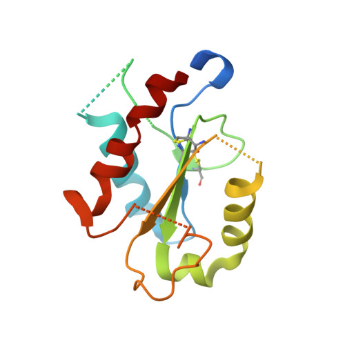 | |
UniProt & NIH Common Fund Data Resources | |||||
PHAROS: P58753 GTEx: ENSG00000150455 | |||||
Entity Groups | |||||
| Sequence Clusters | 30% Identity50% Identity70% Identity90% Identity95% Identity100% Identity | ||||
| UniProt Group | P58753 | ||||
Sequence AnnotationsExpand | |||||
Reference Sequence | |||||
| Length ( Å ) | Angle ( ˚ ) |
|---|---|
| a = 100.253 | α = 90 |
| b = 100.253 | β = 90 |
| c = 78.939 | γ = 90 |
| Software Name | Purpose |
|---|---|
| CBASS | data collection |
| PHASES | phasing |
| CNS | refinement |
| HKL-2000 | data reduction |
| HKL-2000 | data scaling |


