4FZ3 | pdb_00004fz3

Crystal structure of SIRT3 in complex with acetyl p53 peptide coupled with 4-amino-7-methylcoumarin


Experimental Data Snapshot

  • Method: X-RAY DIFFRACTION
  • Resolution: 2.10 Å
  • R-Value Free: 
    0.282 (Depositor), 0.295 (DCC) 
  • R-Value Work: 
    0.208 (Depositor) 
  • R-Value Observed: 
    0.211 (Depositor) 

Starting Model: experimental
View more details

wwPDB Validation 3D Report Full Report

Validation slider image for 4FZ3

This is version 1.3 of the entry. See complete history

Literature

Discovery and Mechanism Study of SIRT1 Activators that Promote the Deacetylation of Fluorophore-Labeled Substrate

Wu, J.Zhang, D.Chen, L.Li, J.Wang, J.Ning, C.Yu, N.Zhao, F.Chen, D.Chen, X.Chen, K.Jiang, H.Liu, H.Liu, D.

(2013) J Med Chem 56: 761-780

  • DOI: https://doi.org/10.1021/jm301032j
  • Primary Citation Related Structures: 
    4FZ3

  • PubMed Abstract: 

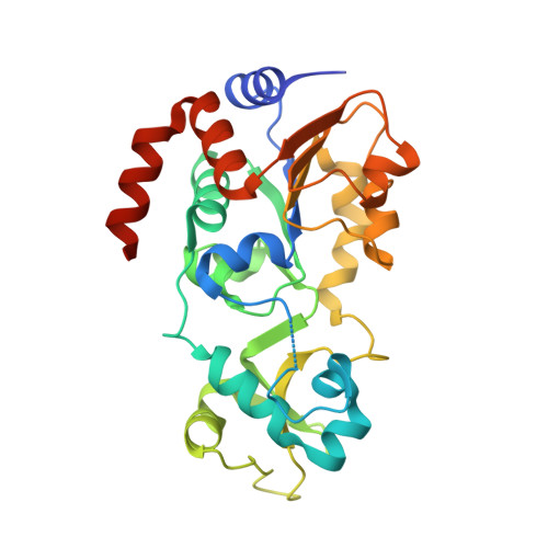
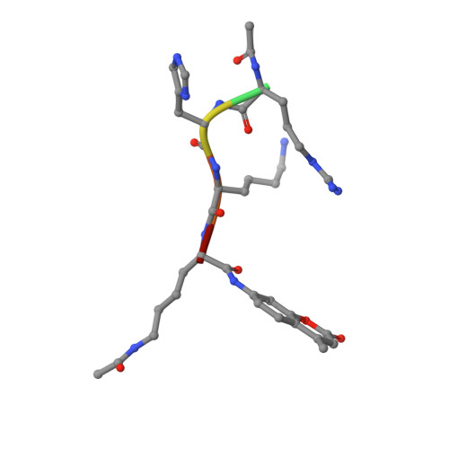
    SIRT1 is an NAD(+)-dependent deacetylase, whose activators have potential therapeutic applications in age-related diseases. Here we report a new class of SIRT1 activators. The activation is dependent on the fluorophore labeled to the substrate. To elucidate the activation mechanism, we solved the crystal structure of SIRT3/ac-RHKK(ac)-AMC complex. The structure revealed that the fluorophore blocked the H-bond formation and created a cavity between the substrate and the Rossmann fold. We built the SIRT1/ac-RHKK(ac)-AMC complex model based on the crystal structure. K(m) and K(d) determinations demonstrated that the fluorophore decreased the peptide binding affinity. The binding modes of SIRT1 activators indicated that a portion of the activators interacts with the fluorophore through π-stacking, while the other portion inserts into the cavity or interacts with the Rossmann fold, thus increasing the substrate affinity. Our study provides new insights into the mechanism of SIRT1 activation and may aid the design of novel SIRT1 activators.


  • Organizational Affiliation
    • Department of Pharmacology III, Shanghai Institute of Materia Medica , Chinese Academy of Sciences, 555 Zu-Chong-Zhi Road, Shanghai 201203, China.

Macromolecule Content 

  • Total Structure Weight: 32.26 kDa 
  • Atom Count: 2,273 
  • Modeled Residue Count: 266 
  • Deposited Residue Count: 289 
  • Unique protein chains: 2

Macromolecules

Find similar proteins by:|  3D Structure
Entity ID: 1
MoleculeChains  Sequence LengthOrganismDetailsImage
NAD-dependent protein deacetylase sirtuin-3, mitochondrial283Homo sapiensMutation(s): 0 
Gene Names: SIRT3
EC: 3.5.1 (PDB Primary Data), 2.3.1 (UniProt), 2.3.1.286 (UniProt)
UniProt & NIH Common Fund Data Resources
Find proteins for Q9NTG7 (Homo sapiens)
Explore Q9NTG7 
Go to UniProtKB:  Q9NTG7
PHAROS:  Q9NTG7
GTEx:  ENSG00000142082 
Entity Groups
Sequence Clusters30% Identity50% Identity70% Identity90% Identity95% Identity100% Identity
UniProt GroupQ9NTG7
Sequence Annotations
Expand
Reference Sequence
Find similar proteins by:  Sequence   |   3D Structure  
Entity ID: 2
MoleculeChains  Sequence LengthOrganismDetailsImage
peptide from Cellular tumor antigen p536Homo sapiensMutation(s): 2 
UniProt & NIH Common Fund Data Resources
Find proteins for P04637 (Homo sapiens)
Explore P04637 
Go to UniProtKB:  P04637
PHAROS:  P04637
GTEx:  ENSG00000141510 
Entity Groups
UniProt GroupP04637
Sequence Annotations
Expand
Reference Sequence

Small Molecules

Ligands 1 Unique
IDChains Name / Formula / InChI Key2D Diagram3D Interactions
ZN

Query on ZN


Download:Ideal Coordinates CCD File
C [auth A]ZINC ION
Zn
PTFCDOFLOPIGGS-UHFFFAOYSA-N
Modified Residues  1 Unique
IDChains TypeFormula2D DiagramParent
ALY
Query on ALY
B
L-PEPTIDE LINKINGC8 H16 N2 O3LYS

Experimental Data & Validation

Experimental Data

  • Method: X-RAY DIFFRACTION
  • Resolution: 2.10 Å
  • R-Value Free:  0.282 (Depositor), 0.295 (DCC) 
  • R-Value Work:  0.208 (Depositor) 
  • R-Value Observed: 0.211 (Depositor) 
Space Group: H 3 2
Unit Cell:
Length ( Å )Angle ( ˚ )
a = 114.809α = 90
b = 114.809β = 90
c = 123.202γ = 120
Software Package:
Software NamePurpose
HKL-2000data collection
MOLREPphasing
REFMACrefinement
HKL-2000data reduction
HKL-2000data scaling

Structure Validation

View Full Validation Report


Entry History 

Deposition Data

Revision History  (Full details and data files)

  • Version 1.0: 2013-03-20
    Type: Initial release
  • Version 1.1: 2023-11-08
    Changes: Data collection, Database references, Derived calculations, Refinement description
  • Version 1.2: 2023-12-06
    Changes: Data collection, Derived calculations
  • Version 1.3: 2024-11-13
    Changes: Structure summary