4FXY

Crystal structure of rat neurolysin with bound pyrazolidin inhibitor


Experimental Data Snapshot

  • Method: X-RAY DIFFRACTION
  • Resolution: 2.80 Å
  • R-Value Free: 0.287 
  • R-Value Work: 0.235 
  • R-Value Observed: 0.240 

wwPDB Validation   3D Report Full Report


Ligand Structure Quality Assessment 


This is version 1.5 of the entry. See complete history


Literature

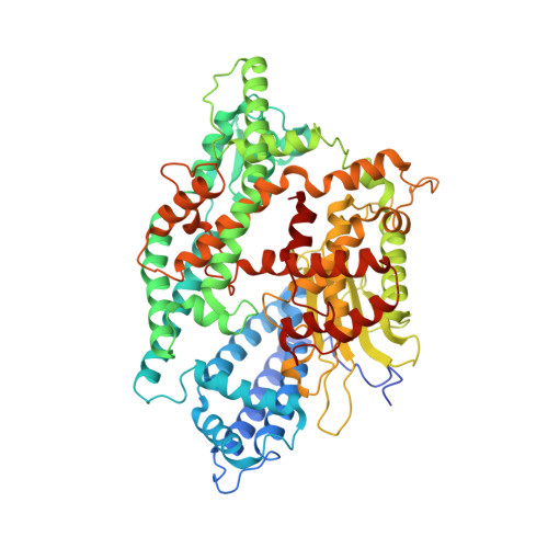
Allosteric inhibition of the neuropeptidase neurolysin.

Hines, C.S.Ray, K.Schmidt, J.J.Xiong, F.Feenstra, R.W.Pras-Raves, M.de Moes, J.P.Lange, J.H.Melikishvili, M.Fried, M.G.Mortenson, P.Charlton, M.Patel, Y.Courtney, S.M.Kruse, C.G.Rodgers, D.W.

(2014) J Biol Chem 289: 35605-35619

  • DOI: https://doi.org/10.1074/jbc.M114.620930
  • Primary Citation of Related Structures:  
    4FXY

  • PubMed Abstract: 

    Neuropeptidases specialize in the hydrolysis of the small bioactive peptides that play a variety of signaling roles in the nervous and endocrine systems. One neuropeptidase, neurolysin, helps control the levels of the dopaminergic circuit modulator neurotensin and is a member of a fold group that includes the antihypertensive target angiotensin converting enzyme. We report the discovery of a potent inhibitor that, unexpectedly, binds away from the enzyme catalytic site. The location of the bound inhibitor suggests it disrupts activity by preventing a hinge-like motion associated with substrate binding and catalysis. In support of this model, the inhibition kinetics are mixed, with both noncompetitive and competitive components, and fluorescence polarization shows directly that the inhibitor reverses a substrate-associated conformational change. This new type of inhibition may have widespread utility in targeting neuropeptidases.


  • Organizational Affiliation

    From the Department of Molecular and Cellular Biochemistry and the Center for Structural Biology, University of Kentucky, Lexington, Kentucky 40536.


Macromolecules
Find similar proteins by:  (by identity cutoff)  |  3D Structure
Entity ID: 1
MoleculeChains Sequence LengthOrganismDetailsImage
Neurolysin, mitochondrialA [auth P],
B [auth Q]
693Rattus norvegicusMutation(s): 1 
Gene Names: Nln
EC: 3.4.24.16
UniProt
Find proteins for P42676 (Rattus norvegicus)
Explore P42676 
Go to UniProtKB:  P42676
Entity Groups  
Sequence Clusters30% Identity50% Identity70% Identity90% Identity95% Identity100% Identity
UniProt GroupP42676
Sequence Annotations
Expand
  • Reference Sequence
Small Molecules
Ligands 2 Unique
IDChains Name / Formula / InChI Key2D Diagram3D Interactions
0W2
Query on 0W2

Download Ideal Coordinates CCD File 
C [auth P],
E [auth Q]
1-{(2S)-1-[(3R)-3-(2-chlorophenyl)-2-(2-fluorophenyl)pyrazolidin-1-yl]-1-oxopropan-2-yl}-3-[(1R,3S,5R,7R)-tricyclo[3.3.1.1~3,7~]dec-2-yl]urea
C29 H34 Cl F N4 O2
LXBLKHIMAYSNHU-XIQYYDOXSA-N
ZN
Query on ZN

Download Ideal Coordinates CCD File 
D [auth P],
F [auth Q]
ZINC ION
Zn
PTFCDOFLOPIGGS-UHFFFAOYSA-N
Experimental Data & Validation

Experimental Data

  • Method: X-RAY DIFFRACTION
  • Resolution: 2.80 Å
  • R-Value Free: 0.287 
  • R-Value Work: 0.235 
  • R-Value Observed: 0.240 
  • Space Group: P 21 21 21
Unit Cell:
Length ( Å )Angle ( ˚ )
a = 88α = 90
b = 131.4β = 90
c = 144.8γ = 90
Software Package:
Software NamePurpose
PHENIXrefinement
CNSrefinement
HKL-2000data collection
HKL-2000data reduction
HKL-2000data scaling
CNSphasing

Structure Validation

View Full Validation Report


Ligand Structure Quality Assessment 


Entry History 

Deposition Data

Revision History  (Full details and data files)

  • Version 1.0: 2013-11-13
    Type: Initial release
  • Version 1.1: 2014-05-07
    Changes: Other
  • Version 1.2: 2014-11-19
    Changes: Database references
  • Version 1.3: 2015-01-14
    Changes: Database references
  • Version 1.4: 2019-07-17
    Changes: Data collection, Refinement description
  • Version 1.5: 2023-09-13
    Changes: Data collection, Database references, Derived calculations, Refinement description