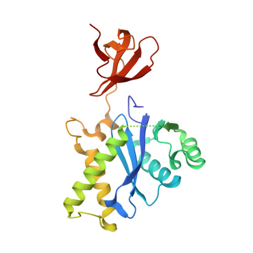
A possible role for the asymmetric C-terminal domain dimer of Rous sarcoma virus integrase in viral DNA binding.
Shi, K., Pandey, K.K., Bera, S., Vora, A.C., Grandgenett, D.P., Aihara, H.(2013) PLoS One 8: e56892-e56892
- PubMed: 23451105 Search on PubMedSearch on PubMed Central
- DOI: https://doi.org/10.1371/journal.pone.0056892
- Primary Citation Related Structures:
4FW1, 4FW2 - PubMed Abstract:
Integration of the retrovirus linear DNA genome into the host chromosome is an essential step in the viral replication cycle, and is catalyzed by the viral integrase (IN). Evidence suggests that IN functions as a dimer that cleaves a dinucleotide from the 3' DNA blunt ends while a dimer of dimers (tetramer) promotes concerted integration of the two processed ends into opposite strands of a target DNA. However, it remains unclear why a dimer rather than a monomer of IN is required for the insertion of each recessed DNA end. To help address this question, we have analyzed crystal structures of the Rous sarcoma virus (RSV) IN mutants complete with all three structural domains as well as its two-domain fragment in a new crystal form at an improved resolution. Combined with earlier structural studies, our results suggest that the RSV IN dimer consists of highly flexible N-terminal domains and a rigid entity formed by the catalytic and C-terminal domains stabilized by the well-conserved catalytic domain dimerization interaction. Biochemical and mutational analyses confirm earlier observations that the catalytic and the C-terminal domains of an RSV IN dimer efficiently integrates one viral DNA end into target DNA. We also show that the asymmetric dimeric interaction between the two C-terminal domains is important for viral DNA binding and subsequent catalysis, including concerted integration. We propose that the asymmetric C-terminal domain dimer serves as a viral DNA binding surface for RSV IN.
- Department of Biochemistry, Molecular Biology and Biophysics, University of Minnesota, Minneapolis, Minnesota, United States of America.
Organizational Affiliation: