Methylated N(omega)-Hydroxy-l-arginine Analogues as Mechanistic Probes for the Second Step of the Nitric Oxide Synthase-Catalyzed Reaction
Jansen Labby, K., Li, H., Roman, L.J., Martasek, P., Poulos, T.L., Silverman, R.B.(2013) Biochemistry 52: 3062-3073
- PubMed: 23586781 Search on PubMedSearch on PubMed Central
- DOI: https://doi.org/10.1021/bi301571v
- Primary Citation Related Structures:
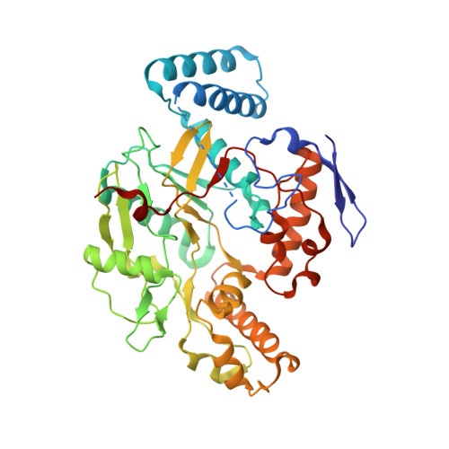
4FVW, 4FVX, 4FVY, 4FVZ, 4FW0, 4GQE - PubMed Abstract:
Nitric oxide synthase (NOS) catalyzes the conversion of L-arginine to L-citrulline through the intermediate N(ω)-hydroxy-L-arginine (NHA), producing nitric oxide, an important mammalian signaling molecule. Several disease states are associated with improper regulation of nitric oxide production, making NOS a therapeutic target. The first step of the NOS reaction has been well-characterized and is presumed to proceed through a compound I heme species, analogous to the cytochrome P450 mechanism. The second step, however, is enzymatically unprecedented and is thought to occur via a ferric peroxo heme species. To gain insight into the details of this unique second step, we report here the synthesis of NHA analogues bearing guanidinium methyl or ethyl substitutions and their investigation as either inhibitors of or alternate substrates for NOS. Radiolabeling studies reveal that N(ω)-methoxy-L-arginine, an alternative NOS substrate, produces citrulline, nitric oxide, and methanol. On the basis of these results, we propose a mechanism for the second step of NOS catalysis in which a methylated nitric oxide species is released and is further metabolized by NOS. Crystal structures of our NHA analogues bound to nNOS have been determined, revealing the presence of an active site water molecule only in the presence of singly methylated analogues. Bulkier analogues displace this active site water molecule; a different mechanism is proposed in the absence of the water molecule. Our results provide new insights into the steric and stereochemical tolerance of the NOS active site and substrate capabilities of NOS.
- Department of Chemistry, Northwestern University, 2145 Sheridan Road, Evanston, IL 60208-3113, USA.
Organizational Affiliation: