4FUS | pdb_00004fus

The X-ray structure of Hahella chejuensis family 48 glycosyl hydrolase


Experimental Data Snapshot

  • Method: X-RAY DIFFRACTION
  • Resolution: 1.75 Å
  • R-Value Free: 
    0.205 (Depositor), 0.209 (DCC) 
  • R-Value Work: 
    0.154 (Depositor), 0.162 (DCC) 
  • R-Value Observed: 
    0.156 (Depositor) 

Starting Model: experimental
View more details

wwPDB Validation 3D Report Full Report

Validation slider image for 4FUS

This is version 2.2 of the entry. See complete history

Literature

Sequence, structure, and evolution of cellulases in glycoside hydrolase family 48.

Sukharnikov, L.O.Alahuhta, M.Brunecky, R.Upadhyay, A.Himmel, M.E.Lunin, V.V.Zhulin, I.B.

(2012) J Biological Chem 287: 41068-41077

  • DOI: https://doi.org/10.1074/jbc.M112.405720
  • Primary Citation Related Structures: 
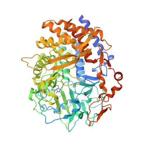
    4FUS

  • PubMed Abstract: 

    Currently, the cost of cellulase enzymes remains a key economic impediment to commercialization of biofuels. Enzymes from glycoside hydrolase family 48 (GH48) are a critical component of numerous natural lignocellulose-degrading systems. Although computational mining of large genomic data sets is a promising new approach for identifying novel cellulolytic activities, current computational methods are unable to distinguish between cellulases and enzymes with different substrate specificities that belong to the same protein family. We show that by using a robust computational approach supported by experimental studies, cellulases and non-cellulases can be effectively identified within a given protein family. Phylogenetic analysis of GH48 showed non-monophyletic distribution, an indication of horizontal gene transfer. Enzymatic function of GH48 proteins coded by horizontally transferred genes was verified experimentally, which confirmed that these proteins are cellulases. Computational and structural studies of GH48 enzymes identified structural elements that define cellulases and can be used to computationally distinguish them from non-cellulases. We propose that the structural element that can be used for in silico discrimination between cellulases and non-cellulases belonging to GH48 is an ω-loop located on the surface of the molecule and characterized by highly conserved rare amino acids. These markers were used to screen metagenomics data for "true" cellulases.


  • Organizational Affiliation
    • BioEnergy Science Center, University of Tennessee, Oak Ridge National Laboratory, Oak Ridge, Tennessee 37831, USA.

Macromolecule Content 

  • Total Structure Weight: 80.08 kDa 
  • Atom Count: 5,837 
  • Modeled Residue Count: 635 
  • Deposited Residue Count: 695 
  • Unique protein chains: 1

Macromolecules

Find similar proteins by:|  3D Structure
Entity ID: 1
MoleculeChains  Sequence LengthOrganismDetailsImage
RTX toxins and related Ca2+-binding protein695Hahella chejuensis KCTC 2396Mutation(s): 0 
Gene Names: HCH_02465
EC: 3.2.1.4
UniProt
Find proteins for Q2SJA2 (Hahella chejuensis (strain KCTC 2396))
Explore Q2SJA2 
Go to UniProtKB:  Q2SJA2
Entity Groups
Sequence Clusters30% Identity50% Identity70% Identity90% Identity95% Identity100% Identity
UniProt GroupQ2SJA2
Sequence Annotations
Expand
Reference Sequence

Oligosaccharides

Help  
Entity ID: 2
MoleculeChains Length2D Diagram GlycosylationD Interactions
beta-D-glucopyranose-(1-4)-beta-D-glucopyranose
B
2N/A
Glycosylation Resources
GlyTouCan: G84824ZO
GlyCosmos: G84824ZO

Small Molecules

Ligands 7 Unique
IDChains Name / Formula / InChI Key2D Diagram3D Interactions
PEG

Query on PEG


Download:Ideal Coordinates CCD File
C [auth A]DI(HYDROXYETHYL)ETHER
C4 H10 O3
MTHSVFCYNBDYFN-UHFFFAOYSA-N
PO4

Query on PO4


Download:Ideal Coordinates CCD File
I [auth A],
J [auth A]
PHOSPHATE ION
O4 P
NBIIXXVUZAFLBC-UHFFFAOYSA-K
GOL

Query on GOL


Download:Ideal Coordinates CCD File
D [auth A],
E [auth A],
F [auth A],
G [auth A],
H [auth A]
GLYCEROL
C3 H8 O3
PEDCQBHIVMGVHV-UHFFFAOYSA-N
EDO

Query on EDO


Download:Ideal Coordinates CCD File
O [auth A]
P [auth A]
Q [auth A]
R [auth A]
S [auth A]
O [auth A],
P [auth A],
Q [auth A],
R [auth A],
S [auth A],
T [auth A],
U [auth A],
V [auth A],
W [auth A],
X [auth A],
Y [auth A],
Z [auth A]
1,2-ETHANEDIOL
C2 H6 O2
LYCAIKOWRPUZTN-UHFFFAOYSA-N
ACT

Query on ACT


Download:Ideal Coordinates CCD File
K [auth A],
L [auth A],
M [auth A],
N [auth A]
ACETATE ION
C2 H3 O2
QTBSBXVTEAMEQO-UHFFFAOYSA-M
CA

Query on CA


Download:Ideal Coordinates CCD File
AA [auth A]CALCIUM ION
Ca
BHPQYMZQTOCNFJ-UHFFFAOYSA-N
NA

Query on NA


Download:Ideal Coordinates CCD File
BA [auth A],
CA [auth A]
SODIUM ION
Na
FKNQFGJONOIPTF-UHFFFAOYSA-N
Modified Residues  1 Unique
IDChains TypeFormula2D DiagramParent
OHI
Query on OHI
A
L-PEPTIDE LINKINGC6 H7 N3 O3HIS

Biologically Interesting Molecules (External Reference) 

1 Unique

Experimental Data & Validation

Experimental Data

  • Method: X-RAY DIFFRACTION
  • Resolution: 1.75 Å
  • R-Value Free:  0.205 (Depositor), 0.209 (DCC) 
  • R-Value Work:  0.154 (Depositor), 0.162 (DCC) 
  • R-Value Observed: 0.156 (Depositor) 
Space Group: P 1 21 1
Unit Cell:
Length ( Å )Angle ( ˚ )
a = 61.69α = 90
b = 78.389β = 98.94
c = 67.075γ = 90
Software Package:
Software NamePurpose
PROTEUM PLUSdata collection
MOLREPphasing
REFMACrefinement
PROTEUM PLUSdata reduction
PROTEUM PLUSdata scaling

Structure Validation

View Full Validation Report


Entry History 

Deposition Data

Revision History  (Full details and data files)

  • Version 1.0: 2012-10-17
    Type: Initial release
  • Version 1.1: 2012-12-19
    Changes: Database references
  • Version 2.0: 2020-07-29
    Type: Remediation
    Reason: Carbohydrate remediation
    Changes: Advisory, Atomic model, Data collection, Database references, Derived calculations, Non-polymer description, Structure summary
  • Version 2.1: 2023-09-13
    Changes: Data collection, Database references, Refinement description, Structure summary
  • Version 2.2: 2023-12-06
    Changes: Data collection