


Crystal Structure of the Urokinase
Kang, Y.N., Stuckey, J.A., Nienaber, V., Giranda, V.To be published.
Experimental Data Snapshot
Entity ID: 1 | |||||
|---|---|---|---|---|---|
| Molecule | Chains | Sequence Length | Organism | Details | Image |
| Urokinase-type plasminogen activator | 246 | Homo sapiens | Mutation(s): 2 Gene Names: PLAU EC: 3.4.21.73 | 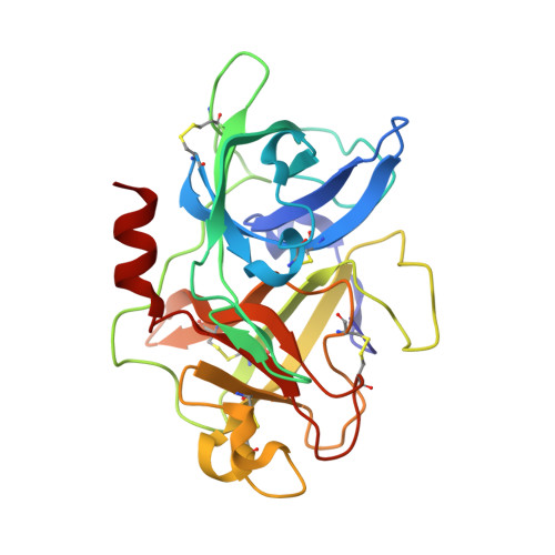 | |
UniProt & NIH Common Fund Data Resources | |||||
PHAROS: P00749 GTEx: ENSG00000122861 | |||||
Entity Groups | |||||
| Sequence Clusters | 30% Identity50% Identity70% Identity90% Identity95% Identity100% Identity | ||||
| UniProt Group | P00749 | ||||
Sequence AnnotationsExpand | |||||
Reference Sequence | |||||
| Ligands 5 Unique | |||||
|---|---|---|---|---|---|
| ID | Chains | Name / Formula / InChI Key | 2D Diagram | 3D Interactions | |
| 7UP Download:Ideal Coordinates CCD File | B [auth A] | 6-(1,2,3,4-tetrahydroisoquinolin-6-ylethynyl)naphthalene-2-carboximidamide C22 H19 N3 ACKRFKIRNILEQJ-UHFFFAOYSA-N |  | ||
| SIN Download:Ideal Coordinates CCD File | C [auth A] | SUCCINIC ACID C4 H6 O4 KDYFGRWQOYBRFD-UHFFFAOYSA-N |  | ||
| SO4 Download:Ideal Coordinates CCD File | D [auth A], E [auth A] | SULFATE ION O4 S QAOWNCQODCNURD-UHFFFAOYSA-L |  | ||
| GOL Download:Ideal Coordinates CCD File | F [auth A], G [auth A], H [auth A] | GLYCEROL C3 H8 O3 PEDCQBHIVMGVHV-UHFFFAOYSA-N |  | ||
| ACT Download:Ideal Coordinates CCD File | I [auth A], J [auth A] | ACETATE ION C2 H3 O2 QTBSBXVTEAMEQO-UHFFFAOYSA-M |  | ||
| Length ( Å ) | Angle ( ˚ ) |
|---|---|
| a = 54.7 | α = 90 |
| b = 53.4 | β = 90 |
| c = 80.25 | γ = 90 |
| Software Name | Purpose |
|---|---|
| BUSTER-TNT | refinement |
| PDB_EXTRACT | data extraction |
| BUSTER | refinement |


