4FQZ | pdb_00004fqz

Crystal structure of a protease-resistant mutant form of human galectin-8


Experimental Data Snapshot

  • Method: X-RAY DIFFRACTION
  • Resolution: 2.80 Å
  • R-Value Free: 
    0.253 (Depositor), 0.246 (DCC) 
  • R-Value Work: 
    0.209 (Depositor), 0.203 (DCC) 
  • R-Value Observed: 
    0.209 (Depositor) 

Starting Model: experimental
View more details

wwPDB Validation 3D Report Full Report

Validation slider image for 4FQZ

This is version 2.1 of the entry. See complete history

Literature

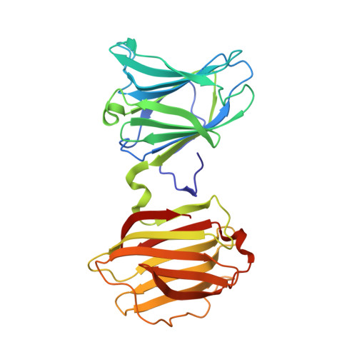
X-ray structure of a protease-resistant mutant form of human galectin-8 with two carbohydrate recognition domains

Yoshida, H.Yamashita, S.Teraoka, M.Itoh, A.Nakakita, S.Nishi, N.Kamitori, S.

(2012) FEBS J 279: 3937-3951

  • DOI: https://doi.org/10.1111/j.1742-4658.2012.08753.x
  • Primary Citation Related Structures: 
    3VKL, 3VKM, 3VKN, 3VKO, 4FQZ

  • PubMed Abstract: 

    Galectin-8 is a tandem-repeat-type β-galactoside-specific animal lectin possessing N-terminal and C-terminal carbohydrate recognition domains (N-CRD and C-CRD, respectively), with a difference in carbohydrate-binding specificity, involved in cell-matrix interaction, malignant transformation, and cell adhesion. N-CRD shows strong affinity for α2-3-sialylated oligosaccharides, a feature unique to galectin-8. C-CRD usually shows lower affinity for oligosaccharides but higher affinity for N-glycan-type branched oligosaccharides than does N-CRD. There have been many structural studies on galectins with a single carbohydrate recognition domain (CRD), but no X-ray structure of a galectin containing both CRDs has been reported. Here, the X-ray structure of a protease-resistant mutant form of human galectin-8 possessing both CRDs and the novel pseudodimer structure of galectin-8 N-CRD in complexes with α2-3-sialylated oligosaccharide ligands were determined. The results revealed a difference in specificity between N-CRD and C-CRD, and provided new insights into the association of CRDs and/or molecules of galectin-8.


  • Organizational Affiliation
    • Life Science Research Center and Faculty of Medicine, Kagawa University, Kagawa, Japan.

Macromolecule Content 

  • Total Structure Weight: 34.07 kDa 
  • Atom Count: 2,438 
  • Modeled Residue Count: 291 
  • Deposited Residue Count: 291 
  • Unique protein chains: 1

Macromolecules

Find similar proteins by:|  3D Structure
Entity ID: 1
MoleculeChains  Sequence LengthOrganismDetailsImage
Galectin-8291Homo sapiensMutation(s): 0 
Gene Names: LGALS8
UniProt & NIH Common Fund Data Resources
Find proteins for O00214 (Homo sapiens)
Explore O00214 
Go to UniProtKB:  O00214
PHAROS:  O00214
GTEx:  ENSG00000116977 
Entity Groups
Sequence Clusters30% Identity50% Identity70% Identity90% Identity95% Identity100% Identity
UniProt GroupO00214
Sequence Annotations
Expand
Reference Sequence

Oligosaccharides

Help  
Entity ID: 2
MoleculeChains Length2D Diagram GlycosylationD Interactions
beta-D-galactopyranose-(1-4)-beta-D-glucopyranose
B
2N/A
Glycosylation Resources
GlyTouCan: G84224TW
GlyCosmos: G84224TW
GlyGen: G84224TW

Small Molecules

Ligands 1 Unique
IDChains Name / Formula / InChI Key2D Diagram3D Interactions
EDO

Query on EDO


Download:Ideal Coordinates CCD File
C [auth A]
D [auth A]
E [auth A]
F [auth A]
G [auth A]
C [auth A],
D [auth A],
E [auth A],
F [auth A],
G [auth A],
H [auth A],
I [auth A],
J [auth A],
K [auth A],
L [auth A]
1,2-ETHANEDIOL
C2 H6 O2
LYCAIKOWRPUZTN-UHFFFAOYSA-N

Biologically Interesting Molecules (External Reference) 

1 Unique

Experimental Data & Validation

Experimental Data

  • Method: X-RAY DIFFRACTION
  • Resolution: 2.80 Å
  • R-Value Free:  0.253 (Depositor), 0.246 (DCC) 
  • R-Value Work:  0.209 (Depositor), 0.203 (DCC) 
  • R-Value Observed: 0.209 (Depositor) 
Space Group: P 43 21 2
Unit Cell:
Length ( Å )Angle ( ˚ )
a = 79.259α = 90
b = 79.259β = 90
c = 129.82γ = 90
Software Package:
Software NamePurpose
ADSCdata collection
MOLREPphasing
CNSrefinement
HKL-2000data reduction
HKL-2000data scaling

Structure Validation

View Full Validation Report


Entry History 

Deposition Data

Revision History  (Full details and data files)

  • Version 1.0: 2012-07-11
    Type: Initial release
  • Version 1.1: 2012-09-12
    Changes: Database references
  • Version 1.2: 2013-07-03
    Changes: Database references
  • Version 1.3: 2017-08-16
    Changes: Refinement description, Source and taxonomy
  • Version 2.0: 2020-07-29
    Type: Remediation
    Reason: Carbohydrate remediation
    Changes: Atomic model, Data collection, Database references, Derived calculations, Structure summary
  • Version 2.1: 2023-11-08
    Changes: Data collection, Database references, Refinement description, Structure summary