Structure-Based Discovery of a Novel Pentamidine-Related Inhibitor of the Calcium-Binding Protein S100B.
McKnight, L.E., Raman, E.P., Bezawada, P., Kudrimoti, S., Wilder, P.T., Hartman, K.G., Godoy-Ruiz, R., Toth, E.A., Coop, A., Mackerell, A.D., Weber, D.J.(2012) ACS Med Chem Lett 3: 975-979
- PubMed: 23264854 Search on PubMedSearch on PubMed Central
- DOI: https://doi.org/10.1021/ml300166s
- Primary Citation Related Structures:
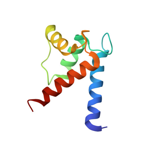
4FQO - PubMed Abstract:
Molecular Dynamics simulations of the pentamidine-S100B complex, where two molecules of pentamidine bind per monomer of S100B, were performed in an effort to determine what properties would be desirable in a pentamidine-derived compound as an inhibitor for S100B. These simulations predicted that increasing the linker length of the compound would allow a single molecule to span both pentamidine binding sites on the protein. The resulting compound, SBi4211 (also known as heptamidine), was synthesized and experiments to study its inhibition of S100B were performed. The 1.65 Å X-ray crystal structure was determined for Ca(2+)-S100B-heptamdine and gives high-resolution information about key contacts that facilitate the interaction between heptamidine and S100B. Additionally, NMR HSQC experiments with both compounds show that heptamidine interacts with the same region of S100B as pentamidine. Heptamidine is able to selectively kill melanoma cells with S100B over those without S100B, indicating that its binding to S100B has an inhibitory effect and that this compound may be useful in designing higher-affinity S100B inhibitors as a treatment for melanoma and other S100B-related cancers.
- Department of Biochemistry and Molecular Biology, University of Maryland School of Medicine, Baltimore, Maryland 21201.
Organizational Affiliation: