4FNC | pdb_00004fnc

Human TDG in a post-reactive complex with 5-hydroxymethyluracil (5hmU)


Experimental Data Snapshot

  • Method: X-RAY DIFFRACTION
  • Resolution: 2.49 Å
  • R-Value Free: 
    0.267 (Depositor), 0.258 (DCC) 
  • R-Value Work: 
    0.224 (Depositor), 0.214 (DCC) 
  • R-Value Observed: 
    0.226 (Depositor) 

Starting Model: experimental
View more details

wwPDB Validation 3D Report Full Report

Validation slider image for 4FNC

This is version 1.4 of the entry. See complete history

Literature

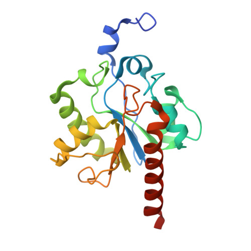
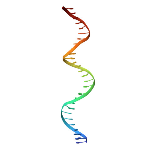
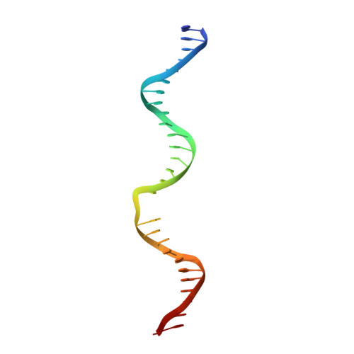
Excision of 5-hydroxymethyluracil and 5-carboxylcytosine by the thymine DNA glycosylase domain: its structural basis and implications for active DNA demethylation.

Hashimoto, H.Hong, S.Bhagwat, A.S.Zhang, X.Cheng, X.

(2012) Nucleic Acids Res 40: 10203-10214

  • DOI: https://doi.org/10.1093/nar/gks845
  • Primary Citation Related Structures: 
    4FNC

  • PubMed Abstract: 

    The mammalian thymine DNA glycosylase (TDG) is implicated in active DNA demethylation via the base excision repair pathway. TDG excises the mismatched base from G:X mismatches, where X is uracil, thymine or 5-hydroxymethyluracil (5hmU). These are, respectively, the deamination products of cytosine, 5-methylcytosine (5mC) and 5-hydroxymethylcytosine (5hmC). In addition, TDG excises the Tet protein products 5-formylcytosine (5fC) and 5-carboxylcytosine (5caC) but not 5hmC and 5mC, when paired with a guanine. Here we present a post-reactive complex structure of the human TDG domain with a 28-base pair DNA containing a G:5hmU mismatch. TDG flips the target nucleotide from the double-stranded DNA, cleaves the N-glycosidic bond and leaves the C1' hydrolyzed abasic sugar in the flipped state. The cleaved 5hmU base remains in a binding pocket of the enzyme. TDG allows hydrogen-bonding interactions to both T/U-based (5hmU) and C-based (5caC) modifications, thus enabling its activity on a wider range of substrates. We further show that the TDG catalytic domain has higher activity for 5caC at a lower pH (5.5) as compared to the activities at higher pH (7.5 and 8.0) and that the structurally related Escherichia coli mismatch uracil glycosylase can excise 5caC as well. We discuss several possible mechanisms, including the amino-imino tautomerization of the substrate base that may explain how TDG discriminates against 5hmC and 5mC.


  • Organizational Affiliation
    • Department of Biochemistry, Emory University School of Medicine, 1510 Clifton Road, Atlanta, GA 30322, USA.

Macromolecule Content 

  • Total Structure Weight: 40.64 kDa 
  • Atom Count: 2,714 
  • Modeled Residue Count: 253 
  • Deposited Residue Count: 261 
  • Unique protein chains: 1
  • Unique nucleic acid chains: 2

Macromolecules


Find similar proteins by:|  3D Structure
Entity ID: 1
MoleculeChains  Sequence LengthOrganismDetailsImage
G/T mismatch-specific thymine DNA glycosylase204Homo sapiensMutation(s): 0 
Gene Names: TDG
EC: 3.2.2.29
UniProt & NIH Common Fund Data Resources
Find proteins for Q13569 (Homo sapiens)
Explore Q13569 
Go to UniProtKB:  Q13569
PHAROS:  Q13569
GTEx:  ENSG00000139372 
Entity Groups
Sequence Clusters30% Identity50% Identity70% Identity90% Identity95% Identity100% Identity
UniProt GroupQ13569
Sequence Annotations
Expand
Reference Sequence
Find similar nucleic acids by:  (by identity cutoff) 
Entity ID: 2
MoleculeChains LengthOrganismImage
DNA (28-MER)B [auth C]28N/A
Sequence Annotations
Expand
Reference Sequence
Find similar nucleic acids by:  (by identity cutoff) 
Entity ID: 3
MoleculeChains LengthOrganismImage
DNA (29-MER)C [auth D]29N/A
Sequence Annotations
Expand
Reference Sequence

Small Molecules

Ligands 1 Unique
IDChains Name / Formula / InChI Key2D Diagram3D Interactions
HMU

Query on HMU


Download:Ideal Coordinates CCD File
D [auth A]5-HYDROXYMETHYL URACIL
C5 H6 N2 O3
JDBGXEHEIRGOBU-UHFFFAOYSA-N

Experimental Data & Validation

Experimental Data

  • Method: X-RAY DIFFRACTION
  • Resolution: 2.49 Å
  • R-Value Free:  0.267 (Depositor), 0.258 (DCC) 
  • R-Value Work:  0.224 (Depositor), 0.214 (DCC) 
  • R-Value Observed: 0.226 (Depositor) 
Space Group: C 1 2 1
Unit Cell:
Length ( Å )Angle ( ˚ )
a = 90.96α = 90
b = 53.72β = 96.49
c = 81.56γ = 90
Software Package:
Software NamePurpose
HKL-2000data collection
PHENIXmodel building
PHENIXrefinement
HKL-2000data reduction
HKL-2000data scaling
PHENIXphasing

Structure Validation

View Full Validation Report


Entry History 

Deposition Data

Revision History  (Full details and data files)

  • Version 1.0: 2012-09-19
    Type: Initial release
  • Version 1.1: 2012-10-17
    Changes: Database references
  • Version 1.2: 2012-11-21
    Changes: Database references
  • Version 1.3: 2014-07-09
    Changes: Non-polymer description
  • Version 1.4: 2023-09-13
    Changes: Advisory, Data collection, Database references, Derived calculations, Refinement description