4FMN

Structure of the C-terminal domain of the Saccharomyces cerevisiae MUTL alpha (MLH1/PMS1) heterodimer bound to a fragment of NTG2


Experimental Data Snapshot

  • Method: X-RAY DIFFRACTION
  • Resolution: 2.69 Å
  • R-Value Free: 0.192 
  • R-Value Work: 0.169 
  • R-Value Observed: 0.170 

wwPDB Validation   3D Report Full Report


This is version 1.3 of the entry. See complete history


Literature

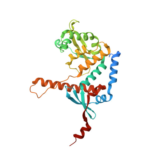
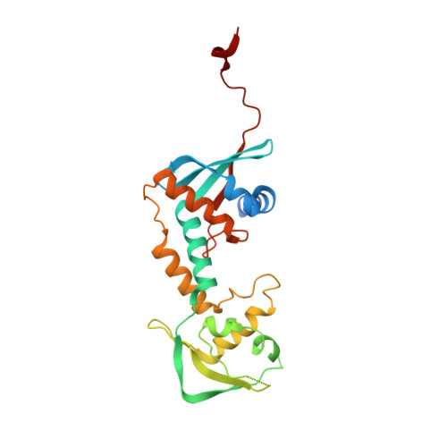
Structure of the MutL alpha C-terminal domain reveals how Mlh1 contributes to Pms1 endonuclease site.

Gueneau, E.Dherin, C.Legrand, P.Tellier-Lebegue, C.Gilquin, B.Bonnesoeur, P.Londino, F.Quemener, C.Le Du, M.H.Marquez, J.A.Moutiez, M.Gondry, M.Boiteux, S.Charbonnier, J.B.

(2013) Nat Struct Mol Biol 20: 461-468

  • DOI: https://doi.org/10.1038/nsmb.2511
  • Primary Citation of Related Structures:  
    4E4W, 4FMN, 4FMO

  • PubMed Abstract: 

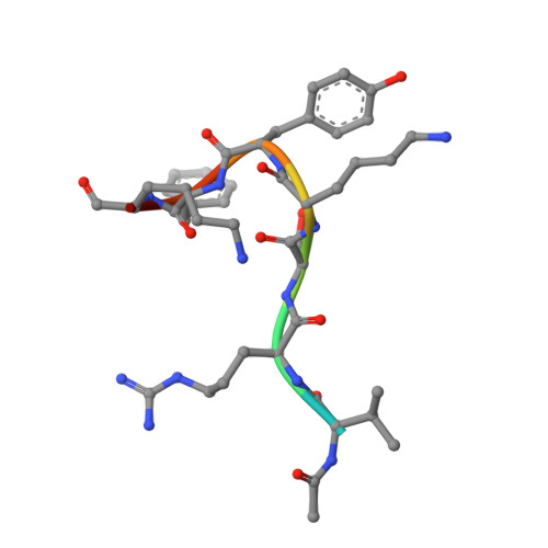
    Mismatch-repair factors have a prominent role in surveying eukaryotic DNA-replication fidelity and in ensuring correct meiotic recombination. These functions depend on MutL-homolog heterodimers with Mlh1. In humans, MLH1 mutations underlie half of hereditary nonpolyposis colorectal cancers (HNPCCs). Here we report crystal structures of the MutLα (Mlh1-Pms1 heterodimer) C-terminal domain (CTD) from Saccharomyces cerevisiae, alone and in complex with fragments derived from Mlh1 partners. These structures reveal structural rearrangements and additional domains in MutLα as compared to the bacterial MutL counterparts and show that the strictly conserved C terminus of Mlh1 forms part of the Pms1 endonuclease site. The structures of the ternary complexes between MutLα(CTD) and Exo1 or Ntg2 fragments reveal the binding mode of the MIP-box motif shared by several Mlh1 partners. Finally, the structures provide a rationale for the deleterious impact of MLH1 mutations in HNPCCs.


  • Organizational Affiliation

    Unité Mixte de Recherche 8221, Commissariat à l'Energie Atomique, Centre National de la Recherche Scientifique, Institut de Biologie et Technologies de Saclay, Gif-sur-Yvette, France.


Macromolecules
Find similar proteins by:  (by identity cutoff)  |  3D Structure
Entity ID: 1
MoleculeChains Sequence LengthOrganismDetailsImage
DNA mismatch repair protein MLH1288Saccharomyces cerevisiae S288CMutation(s): 0 
Gene Names: MLH1PMS2YMR167WYM8520.16
UniProt
Find proteins for P38920 (Saccharomyces cerevisiae (strain ATCC 204508 / S288c))
Explore P38920 
Go to UniProtKB:  P38920
Entity Groups  
Sequence Clusters30% Identity50% Identity70% Identity90% Identity95% Identity100% Identity
UniProt GroupP38920
Sequence Annotations
Expand
  • Reference Sequence
Find similar proteins by:  (by identity cutoff)  |  3D Structure
Entity ID: 2
MoleculeChains Sequence LengthOrganismDetailsImage
DNA mismatch repair protein PMS1240Saccharomyces cerevisiae S288CMutation(s): 0 
Gene Names: PMS1YNL082WN2317
UniProt
Find proteins for P14242 (Saccharomyces cerevisiae (strain ATCC 204508 / S288c))
Explore P14242 
Go to UniProtKB:  P14242
Entity Groups  
Sequence Clusters30% Identity50% Identity70% Identity90% Identity95% Identity100% Identity
UniProt GroupP14242
Sequence Annotations
Expand
  • Reference Sequence

Find similar proteins by:  Sequence   |   3D Structure  

Entity ID: 3
MoleculeChains Sequence LengthOrganismDetailsImage
DNA repair peptide9Saccharomyces cerevisiae S288CMutation(s): 1 
UniProt
Find proteins for Q08214 (Saccharomyces cerevisiae (strain ATCC 204508 / S288c))
Explore Q08214 
Go to UniProtKB:  Q08214
Entity Groups  
UniProt GroupQ08214
Sequence Annotations
Expand
  • Reference Sequence
Small Molecules
Ligands 3 Unique
IDChains Name / Formula / InChI Key2D Diagram3D Interactions
GOL
Query on GOL

Download Ideal Coordinates CCD File 
D [auth A],
E [auth A],
F [auth A],
G [auth A],
H [auth A]
GLYCEROL
C3 H8 O3
PEDCQBHIVMGVHV-UHFFFAOYSA-N
ZN
Query on ZN

Download Ideal Coordinates CCD File 
N [auth B],
O [auth B]
ZINC ION
Zn
PTFCDOFLOPIGGS-UHFFFAOYSA-N
EDO
Query on EDO

Download Ideal Coordinates CCD File 
I [auth A]
J [auth A]
K [auth A]
L [auth A]
M [auth A]
I [auth A],
J [auth A],
K [auth A],
L [auth A],
M [auth A],
P [auth B]
1,2-ETHANEDIOL
C2 H6 O2
LYCAIKOWRPUZTN-UHFFFAOYSA-N
Experimental Data & Validation

Experimental Data

  • Method: X-RAY DIFFRACTION
  • Resolution: 2.69 Å
  • R-Value Free: 0.192 
  • R-Value Work: 0.169 
  • R-Value Observed: 0.170 
  • Space Group: C 1 2 1
Unit Cell:
Length ( Å )Angle ( ˚ )
a = 190.94α = 90
b = 66.3β = 90.15
c = 74.11γ = 90
Software Package:
Software NamePurpose
MOLREPphasing
BUSTERrefinement
XDSdata reduction
XDSdata scaling

Structure Validation

View Full Validation Report


Entry History 

Deposition Data

Revision History  (Full details and data files)

  • Version 1.0: 2013-02-20
    Type: Initial release
  • Version 1.1: 2013-02-27
    Changes: Structure summary
  • Version 1.2: 2013-08-28
    Changes: Database references
  • Version 1.3: 2023-09-13
    Changes: Data collection, Database references, Derived calculations, Refinement description