4FGZ

Crystal Structure of Phosphoethanolamine Methyltransferase from Plasmodium falciparum in Complex with Amodiaquine


Experimental Data Snapshot

  • Method: X-RAY DIFFRACTION
  • Resolution: 1.99 Å
  • R-Value Free: 0.227 
  • R-Value Work: 0.181 
  • R-Value Observed: 0.183 

wwPDB Validation   3D Report Full Report


Ligand Structure Quality Assessment 


This is version 1.1 of the entry. See complete history


Literature

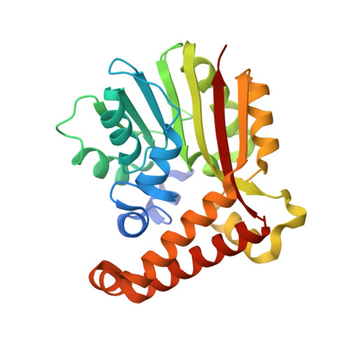
Crystal structure of phosphoethanolamine methyltransferase from Plasmodium falciparum in complex with amodiaquine.

Lee, S.G.Alpert, T.D.Jez, J.M.

(2012) Bioorg Med Chem Lett 22: 4990-4993

  • DOI: https://doi.org/10.1016/j.bmcl.2012.06.032
  • Primary Citation of Related Structures:  
    4FGZ

  • PubMed Abstract: 
  • Phosphoethanolamine N-methyltransferase (PMT) is essential for phospholipid biogenesis in the malarial parasite Plasmodium falciparum. PfPMT catalyzes the triple methylation of phosphoethanolamine to produce phosphocholine, which is then used for phosphatidylcholine synthesis ...

    Phosphoethanolamine N-methyltransferase (PMT) is essential for phospholipid biogenesis in the malarial parasite Plasmodium falciparum. PfPMT catalyzes the triple methylation of phosphoethanolamine to produce phosphocholine, which is then used for phosphatidylcholine synthesis. Here we describe the 2.0Å resolution X-ray crystal structure of PfPMT in complex with amodiaquine. To better characterize inhibition of PfPMT by amodiaquine, we determined the IC(50) values of a series of aminoquinolines using a direct radiochemical assay. Both structural and functional analyses provide a possible approach for the development of new small molecule inhibitors of PfPMT.


    Organizational Affiliation

    Department of Biology, Washington University, St. Louis, MO 63130, USA.


Macromolecules
Find similar proteins by:  (by identity cutoff)  |  3D Structure
Entity ID: 1
MoleculeChainsSequence LengthOrganismDetailsImage
Phosphoethanolamine N-methyltransferase
A, B
266Plasmodium falciparumMutation(s): 0 
Gene Names: PMT
EC: 2.1.1.103
UniProt
Find proteins for Q6T755 (Plasmodium falciparum)
Explore Q6T755 
Go to UniProtKB:  Q6T755
Entity Groups  
Sequence Clusters30% Identity50% Identity70% Identity90% Identity95% Identity100% Identity
UniProt GroupQ6T755
Protein Feature View
Expand
  • Reference Sequence
Small Molecules
Ligands 3 Unique
IDChainsName / Formula / InChI Key2D Diagram3D Interactions
SAH
Query on SAH

Download Ideal Coordinates CCD File 
E [auth A],
I [auth B]
S-ADENOSYL-L-HOMOCYSTEINE
C14 H20 N6 O5 S
ZJUKTBDSGOFHSH-WFMPWKQPSA-N
 Ligand Interaction
CQA
Query on CQA

Download Ideal Coordinates CCD File 
C [auth A],
D [auth A],
G [auth B],
H [auth B]
4-[(7-CHLOROQUINOLIN-4-YL)AMINO]-2-[(DIETHYLAMINO)METHYL]PHENOL
C20 H22 Cl N3 O
OVCDSSHSILBFBN-UHFFFAOYSA-N
 Ligand Interaction
PO4
Query on PO4

Download Ideal Coordinates CCD File 
F [auth A],
J [auth B]
PHOSPHATE ION
O4 P
NBIIXXVUZAFLBC-UHFFFAOYSA-K
 Ligand Interaction
Binding Affinity Annotations 
IDSourceBinding Affinity
CQA Binding MOAD:  4FGZ IC50: 1.35e+6 (nM) from 1 assay(s)
PDBBind:  4FGZ IC50: 1.35e+6 (nM) from 1 assay(s)
BindingDB:  4FGZ IC50: 1.35e+6 (nM) from 1 assay(s)
Experimental Data & Validation

Experimental Data

  • Method: X-RAY DIFFRACTION
  • Resolution: 1.99 Å
  • R-Value Free: 0.227 
  • R-Value Work: 0.181 
  • R-Value Observed: 0.183 
  • Space Group: P 1 2 1
Unit Cell:
Length ( Å )Angle ( ˚ )
a = 66.051α = 90
b = 60.639β = 90.008
c = 68.151γ = 90
Software Package:
Software NamePurpose
HKL-3000data collection
PHASERphasing
PHENIXrefinement
HKL-3000data reduction
HKL-3000data scaling

Structure Validation

View Full Validation Report


Ligand Structure Quality Assessment 


Entry History 

Deposition Data

Revision History  (Full details and data files)

  • Version 1.0: 2012-07-25
    Type: Initial release
  • Version 1.1: 2012-08-01
    Changes: Database references