4FER

Crystal structure of Bacillus Subtilis expansin (EXLX1) in complex with cellohexaose


Experimental Data Snapshot

  • Method: X-RAY DIFFRACTION
  • Resolution: 2.10 Å
  • R-Value Free: 0.205 
  • R-Value Work: 0.167 
  • R-Value Observed: 0.169 

wwPDB Validation   3D Report Full Report


This is version 2.1 of the entry. See complete history


Literature

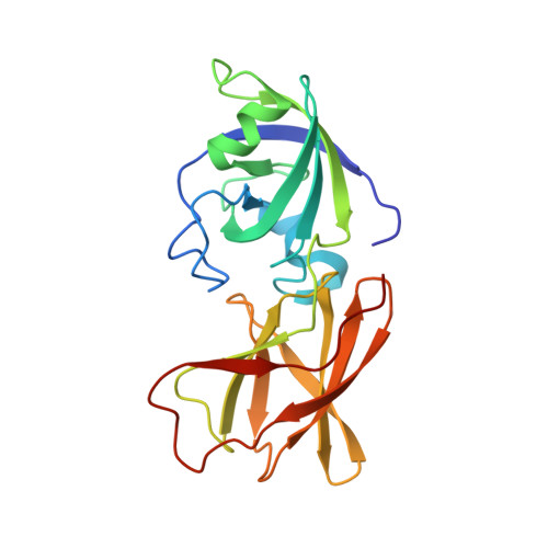
Structural basis for entropy-driven cellulose binding by a type-A cellulose-binding module (CBM) and bacterial expansin.

Georgelis, N.Yennawar, N.H.Cosgrove, D.J.

(2012) Proc Natl Acad Sci U S A 109: 14830-14835

  • DOI: https://doi.org/10.1073/pnas.1213200109
  • Primary Citation of Related Structures:  
    4FER, 4FFT, 4FG2, 4FG4

  • PubMed Abstract: 

    Components of modular cellulases, type-A cellulose-binding modules (CBMs) bind to crystalline cellulose and enhance enzyme effectiveness, but structural details of the interaction are uncertain. We analyzed cellulose binding by EXLX1, a bacterial expansin with ability to loosen plant cell walls and whose domain D2 has type-A CBM characteristics. EXLX1 strongly binds to crystalline cellulose via D2, whereas its affinity for soluble cellooligosaccharides is weak. Calorimetry indicated cellulose binding was largely entropically driven. We solved the crystal structures of EXLX1 complexed with cellulose-like oligosaccharides to find that EXLX1 binds the ligands through hydrophobic interactions of three linearly arranged aromatic residues in D2. The crystal structures revealed a unique form of ligand-mediated dimerization, with the oligosaccharide sandwiched between two D2 domains in opposite polarity. This report clarifies the molecular target of expansin and the specific molecular interactions of a type-A CBM with cellulose.


  • Organizational Affiliation

    Department of Biology, Pennsylvania State University, University Park, PA 16802, USA.


Macromolecules
Find similar proteins by:  (by identity cutoff)  |  3D Structure
Entity ID: 1
MoleculeChains Sequence LengthOrganismDetailsImage
Expansin-yoaJ
A, B
208Bacillus subtilis subsp. subtilis str. 168Mutation(s): 0 
Gene Names: yoaJBSU18630
UniProt
Find proteins for O34918 (Bacillus subtilis (strain 168))
Explore O34918 
Go to UniProtKB:  O34918
Entity Groups  
Sequence Clusters30% Identity50% Identity70% Identity90% Identity95% Identity100% Identity
UniProt GroupO34918
Sequence Annotations
Expand
  • Reference Sequence
Oligosaccharides

Help

Entity ID: 2
MoleculeChains Length2D Diagram Glycosylation3D Interactions
beta-D-glucopyranose-(1-4)-beta-D-glucopyranose-(1-4)-beta-D-glucopyranose-(1-4)-beta-D-glucopyranose-(1-4)-beta-D-glucopyranose-(1-4)-beta-D-glucopyranose
C
6N/A
Glycosylation Resources
GlyTouCan:  G09454VW
GlyCosmos:  G09454VW
GlyGen:  G09454VW
Biologically Interesting Molecules (External Reference) 1 Unique
Experimental Data & Validation

Experimental Data

  • Method: X-RAY DIFFRACTION
  • Resolution: 2.10 Å
  • R-Value Free: 0.205 
  • R-Value Work: 0.167 
  • R-Value Observed: 0.169 
  • Space Group: P 31
Unit Cell:
Length ( Å )Angle ( ˚ )
a = 58.205α = 90
b = 58.205β = 90
c = 147.277γ = 120
Software Package:
Software NamePurpose
CrystalCleardata collection
PHENIXmodel building
PHENIXrefinement
HKL-2000data reduction
HKL-2000data scaling
PHENIXphasing

Structure Validation

View Full Validation Report


Entry History 

Deposition Data

Revision History  (Full details and data files)

  • Version 1.0: 2012-08-22
    Type: Initial release
  • Version 1.1: 2012-09-26
    Changes: Database references
  • Version 1.2: 2015-06-24
    Changes: Derived calculations
  • Version 2.0: 2020-07-29
    Type: Remediation
    Reason: Carbohydrate remediation
    Changes: Atomic model, Data collection, Database references, Derived calculations, Non-polymer description, Structure summary
  • Version 2.1: 2024-02-28
    Changes: Data collection, Database references, Structure summary