4FEO

Crystal structure of the AU25A/A46G/C74U mutant xpt-pbuX guanine riboswitch aptamer domain in complex with 2,6-diaminopurine


Experimental Data Snapshot

  • Method: X-RAY DIFFRACTION
  • Resolution: 1.60 Å
  • R-Value Free: 0.228 
  • R-Value Work: 0.174 
  • R-Value Observed: 0.174 

wwPDB Validation   3D Report Full Report


Ligand Structure Quality Assessment 


This is version 1.3 of the entry. See complete history


Literature

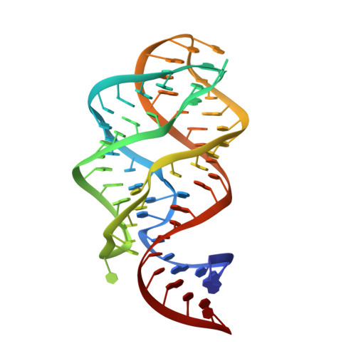
Nucleotides Adjacent to the Ligand-Binding Pocket are Linked to Activity Tuning in the Purine Riboswitch.

Stoddard, C.D.Widmann, J.Trausch, J.J.Marcano-Velazquez, J.G.Knight, R.Batey, R.T.

(2013) J Mol Biol 425: 1596-1611

  • DOI: https://doi.org/10.1016/j.jmb.2013.02.023
  • Primary Citation of Related Structures:  
    4FEJ, 4FEL, 4FEN, 4FEO, 4FEP

  • PubMed Abstract: 

    Direct sensing of intracellular metabolite concentrations by riboswitch RNAs provides an economical and rapid means to maintain metabolic homeostasis. Since many organisms employ the same class of riboswitch to control different genes or transcription units, it is likely that functional variation exists in riboswitches such that activity is tuned to meet cellular needs. Using a bioinformatic approach, we have identified a region of the purine riboswitch aptamer domain that displays conservation patterns linked to riboswitch activity. Aptamer domain compositions within this region can be divided into nine classes that display a spectrum of activities. Naturally occurring compositions in this region favor rapid association rate constants and slow dissociation rate constants for ligand binding. Using X-ray crystallography and chemical probing, we demonstrate that both the free and bound states are influenced by the composition of this region and that modest sequence alterations have a dramatic impact on activity. The introduction of non-natural compositions result in the inability to regulate gene expression in vivo, suggesting that aptamer domain activity is highly plastic and thus readily tunable to meet cellular needs.


  • Organizational Affiliation

    Department of Chemistry and Biochemistry, 596 UCB, University of Colorado, Boulder, CO 80309-0596, USA.


Macromolecules
Find similar nucleic acids by:  (by identity cutoff)  |  3D Structure
Entity ID: 1
MoleculeChains LengthOrganismImage
U25A/A46G/C74U mutant of the B. subtilis xpt-pbuX guanine riboswitch aptamer domainA [auth B]67N/A
Sequence Annotations
Expand
  • Reference Sequence
Small Molecules
Ligands 2 Unique
IDChains Name / Formula / InChI Key2D Diagram3D Interactions
NCO
Query on NCO

Download Ideal Coordinates CCD File 
C [auth B]
D [auth B]
E [auth B]
F [auth B]
G [auth B]
C [auth B],
D [auth B],
E [auth B],
F [auth B],
G [auth B],
H [auth B],
I [auth B],
J [auth B],
K [auth B]
COBALT HEXAMMINE(III)
Co H18 N6
DYLMFCCYOUSRTK-UHFFFAOYSA-N
6AP
Query on 6AP

Download Ideal Coordinates CCD File 
B
9H-PURINE-2,6-DIAMINE
C5 H6 N6
MSSXOMSJDRHRMC-UHFFFAOYSA-N
Experimental Data & Validation

Experimental Data

  • Method: X-RAY DIFFRACTION
  • Resolution: 1.60 Å
  • R-Value Free: 0.228 
  • R-Value Work: 0.174 
  • R-Value Observed: 0.174 
  • Space Group: C 1 2 1
Unit Cell:
Length ( Å )Angle ( ˚ )
a = 132.91α = 90
b = 35.23β = 90.36
c = 41.46γ = 90
Software Package:
Software NamePurpose
CrystalCleardata collection
CNSrefinement
CrystalCleardata reduction
CrystalCleardata scaling
CNSphasing

Structure Validation

View Full Validation Report


Ligand Structure Quality Assessment 


Entry History 

Deposition Data

Revision History  (Full details and data files)

  • Version 1.0: 2013-02-27
    Type: Initial release
  • Version 1.1: 2013-03-27
    Changes: Database references
  • Version 1.2: 2013-05-22
    Changes: Database references
  • Version 1.3: 2024-02-28
    Changes: Data collection, Database references, Derived calculations