4FEE

High-resolution structure of pyruvate oxidase in complex with reaction intermediate 2-hydroxyethyl-thiamin diphosphate carbanion-enamine, crystal B


Experimental Data Snapshot

  • Method: X-RAY DIFFRACTION
  • Resolution: 1.13 Å
  • R-Value Free: 0.149 
  • R-Value Work: 0.122 

wwPDB Validation   3D Report Full Report


Ligand Structure Quality Assessment 


This is version 2.0 of the entry. See complete history


Literature

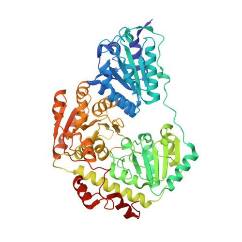
Unexpected tautomeric equilibria of the carbanion-enamine intermediate in pyruvate oxidase highlight unrecognized chemical versatility of thiamin.

Meyer, D.Neumann, P.Koers, E.Sjuts, H.Ludtke, S.Sheldrick, G.M.Ficner, R.Tittmann, K.

(2012) Proc Natl Acad Sci U S A 109: 10867-10872

  • DOI: https://doi.org/10.1073/pnas.1201280109
  • Primary Citation of Related Structures:  
    4FEE, 4FEG

  • PubMed Abstract: 

    Thiamin diphosphate, the vitamin B1 coenzyme, plays critical roles in fundamental metabolic pathways that require acyl carbanion equivalents. Studies on chemical models and enzymes had suggested that these carbanions are resonance-stabilized as enamines. A crystal structure of this intermediate in pyruvate oxidase at 1.1 Å resolution now challenges this paradigm by revealing that the enamine does not accumulate. Instead, the intermediate samples between the ketone and the carbanion both interlocked in a tautomeric equilibrium. Formation of the keto tautomer is associated with a loss of aromaticity of the cofactor. The alternate confinement of electrons to neighboring atoms rather than π-conjugation seems to be of importance for the enzyme-catalyzed, redox-coupled acyl transfer to phosphate, which requires a dramatic inversion of polarity of the reacting substrate carbon in two subsequent catalytic steps. The ability to oscillate between a nucleophilic (carbanion) and an electrophilic (ketone) substrate center highlights a hitherto unrecognized versatility of the thiamin cofactor. It remains to be studied whether formation of the keto tautomer is a general feature of all thiamin enzymes, as it could provide for stable storage of the carbanion state, or whether this feature represents a specific trait of thiamin oxidases. In addition, the protonation state of the two-electron reduced flavin cofactor can be fully assigned, demonstrating the power of high-resolution cryocrystallography for elucidation of enzymatic mechanisms.


  • Organizational Affiliation

    Albrecht-von-Haller Institute and Göttingen Center for Molecular Biosciences, D-37077 Göttingen, Germany.


Macromolecules
Find similar proteins by:  (by identity cutoff)  |  3D Structure
Entity ID: 1
MoleculeChains Sequence LengthOrganismDetailsImage
Pyruvate oxidase
A, B
603Lactiplantibacillus plantarum JDM1Mutation(s): 0 
Gene Names: pox5JDM1_2870
EC: 1.2.3.3
Entity Groups  
Sequence Clusters30% Identity50% Identity70% Identity90% Identity95% Identity100% Identity
Sequence Annotations
Expand
  • Reference Sequence
Small Molecules
Ligands 6 Unique
IDChains Name / Formula / InChI Key2D Diagram3D Interactions
FAD
Query on FAD

Download Ideal Coordinates CCD File 
C [auth A],
M [auth B]
FLAVIN-ADENINE DINUCLEOTIDE
C27 H33 N9 O15 P2
VWWQXMAJTJZDQX-UYBVJOGSSA-N
TDM
Query on TDM

Download Ideal Coordinates CCD File 
J [auth A],
P [auth B]
2-[(2E)-3-[(4-AMINO-2-METHYLPYRIMIDIN-5-YL)METHYL]-2-(1-HYDROXYETHYLIDENE)-4-METHYL-2,3-DIHYDRO-1,3-THIAZOL-5-YL]ETHYL TRIHYDROGEN DIPHOSPHATE
C14 H22 N4 O8 P2 S
ZZLCKFKIIVXBQU-NTEUORMPSA-N
PO4
Query on PO4

Download Ideal Coordinates CCD File 
I [auth A],
Q [auth B]
PHOSPHATE ION
O4 P
NBIIXXVUZAFLBC-UHFFFAOYSA-K
GOL
Query on GOL

Download Ideal Coordinates CCD File 
D [auth A]
G [auth A]
H [auth A]
K [auth A]
R [auth B]
D [auth A],
G [auth A],
H [auth A],
K [auth A],
R [auth B],
S [auth B]
GLYCEROL
C3 H8 O3
PEDCQBHIVMGVHV-UHFFFAOYSA-N
PYR
Query on PYR

Download Ideal Coordinates CCD File 
E [auth A],
F [auth A],
N [auth B],
O [auth B]
PYRUVIC ACID
C3 H4 O3
LCTONWCANYUPML-UHFFFAOYSA-N
MG
Query on MG

Download Ideal Coordinates CCD File 
L [auth A],
T [auth B]
MAGNESIUM ION
Mg
JLVVSXFLKOJNIY-UHFFFAOYSA-N
Experimental Data & Validation

Experimental Data

  • Method: X-RAY DIFFRACTION
  • Resolution: 1.13 Å
  • R-Value Free: 0.149 
  • R-Value Work: 0.122 
  • Space Group: C 2 2 21
Unit Cell:
Length ( Å )Angle ( ˚ )
a = 119.07α = 90
b = 154.46β = 90
c = 165.82γ = 90
Software Package:
Software NamePurpose
XSCALEdata scaling
SHELXrefinement
PDB_EXTRACTdata extraction
DNAdata collection
XDSdata reduction
PHENIXphasing
SHELXL-97refinement

Structure Validation

View Full Validation Report


Ligand Structure Quality Assessment 


Entry History 

Deposition Data

Revision History  (Full details and data files)

  • Version 1.0: 2012-06-20
    Type: Initial release
  • Version 1.1: 2012-07-11
    Changes: Database references
  • Version 1.2: 2012-07-25
    Changes: Database references
  • Version 1.3: 2023-09-13
    Changes: Data collection, Database references, Derived calculations, Refinement description
  • Version 2.0: 2023-11-15
    Changes: Atomic model, Data collection