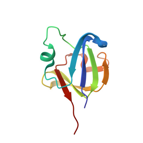
Aggregation-prone near-native intermediate formation during unfolding of a structurally similar nonlenticular beta/gamma-crystallin domain
Rajanikanth, V., Srivastava, S.S., Singh, A.K., Rajyalakshmi, M., Chandra, K., Aravind, P., Sankaranarayanan, R., Sharma, Y.(2012) Biochemistry 51: 8502-8513
- PubMed: 23043265 Search on PubMed
- DOI: https://doi.org/10.1021/bi300844u
- Primary Citation Related Structures:
4FD9 - PubMed Abstract:
The folding and unfolding of structurally similar proteins belonging to a family have long been a focus of investigation of the structure-(un)folding relationship. Such studies are yet to reach a consensus about whether structurally similar domains follow common or different unfolding pathways. Members of the βγ-crystallin superfamily, which consists of structurally similar proteins with limited sequence similarity from diverse life forms spanning microbes to mammals, form an appropriate model system for exploring this relationship further. We selected a new member, Crybg3_D3, the third βγ-crystallin domain of non-lens vertebrate protein Crybg3 from mouse brain. The crystal structure determined at 1.86 Å demonstrates that the βγ-crystallin domain of Crybg3 resembles more closely the lens βγ-crystallins than the microbial crystallins do. However, interestingly, this structural cousin follows a quite distinct (un)folding pathway via formation of an intermediate state. The intermediate species is in a nativelike conformation with variation in flexibility and tends to form insoluble aggregates. The individual domains of lens βγ-crystallins (and microbial homologues) do not follow such an unfolding pattern. Thus, even the closest members of a subfamily within a superfamily do not necessarily follow similar unfolding paths, suggesting the divergence acquired by these domains, which could be observed only by unfolding. Additionally, this study provides insights into the modifications that this domain has undergone during its recruitment into the non-lens tissues in vertebrates.
- Centre for Cellular and Molecular Biology (CCMB), Council of Scientific and Industrial Research (CSIR), Uppal Road, Hyderabad 500 007, India.
Organizational Affiliation: