4F7H | pdb_00004f7h

The crystal structure of kindlin-2 pleckstrin homology domain in free form


Experimental Data Snapshot

  • Method: X-RAY DIFFRACTION
  • Resolution: 1.90 Å
  • R-Value Free: 
    0.224 (Depositor), 0.223 (DCC) 
  • R-Value Work: 
    0.195 (Depositor), 0.196 (DCC) 
  • R-Value Observed: 
    0.196 (Depositor) 

Starting Model: experimental
View more details

wwPDB Validation 3D Report Full Report

Validation slider image for 4F7H

Ligand Structure Quality Assessment 


This is version 1.2 of the entry. See complete history

Literature

Crystal structure of kindlin-2 PH domain reveals a conformational transition for its membrane anchoring and regulation of integrin activation.

Liu, Y.Zhu, Y.Ye, S.Zhang, R.

(2012) Protein Cell 3: 434-440

  • DOI: https://doi.org/10.1007/s13238-012-2046-1
  • Primary Citation Related Structures: 
    4F7H

  • PubMed Abstract: 

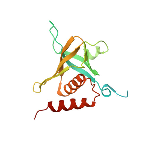
    Kindlin-2 belongs to a subfamily of FERM domain containing proteins, which plays key roles in activating integrin transmembrane receptors and mediating cell adhesion. Compared to conventional FERM domains, kindlin-2 FERM contains an inserted pleckstrin homology (PH) domain that specifically binds to phosphatidylinositol (3,4,5) trisphosphate (PIP3) and regulates the kindlin-2 function. We have determined the crystal structure of kindlin-2 PH domain at 1.9 Å resolution, which reveals a conserved PH domain fold with a highly charged and open binding pocket for PIP3 head group. Structural comparison with a previously reported solution structure of kindlin-2 PH domain bound to PIP3 head group reveals that upon PIP3 insertion, there is a significant conformational change of both the highly positively charged loop at the entry of the PIP3 binding pocket and the entire β barrel of the PH domain. We propose that such "induced-fit" type change is crucial for the tight binding of PIP3 to anchor kindlin-2 onto the membrane surface, thereby promoting its binding to integrins. Our results provide important structural insight into kindlin-2-mediated membrane anchoring and integrin activation.


  • Organizational Affiliation
    • National Laboratory of Biophysics, Institute of Biophysics, Chinese Academy of Sciences, Beijing, China.

Macromolecule Content 

  • Total Structure Weight: 19.73 kDa 
  • Atom Count: 1,182 
  • Modeled Residue Count: 135 
  • Deposited Residue Count: 173 
  • Unique protein chains: 1

Macromolecules

Find similar proteins by:|  3D Structure
Entity ID: 1
MoleculeChains  Sequence LengthOrganismDetailsImage
Fermitin family homolog 2173Homo sapiensMutation(s): 0 
Gene Names: FERMT2KIND2MIG2PLEKHC1
Membrane Entity: Yes 
UniProt & NIH Common Fund Data Resources
Find proteins for Q96AC1 (Homo sapiens)
Explore Q96AC1 
Go to UniProtKB:  Q96AC1
PHAROS:  Q96AC1
GTEx:  ENSG00000073712 
Entity Groups
Sequence Clusters30% Identity50% Identity70% Identity90% Identity95% Identity100% Identity
UniProt GroupQ96AC1
Sequence Annotations
Expand
Reference Sequence

Experimental Data & Validation

Experimental Data

  • Method: X-RAY DIFFRACTION
  • Resolution: 1.90 Å
  • R-Value Free:  0.224 (Depositor), 0.223 (DCC) 
  • R-Value Work:  0.195 (Depositor), 0.196 (DCC) 
  • R-Value Observed: 0.196 (Depositor) 
Space Group: P 43 21 2
Unit Cell:
Length ( Å )Angle ( ˚ )
a = 58.474α = 90
b = 58.474β = 90
c = 92.249γ = 90
Software Package:
Software NamePurpose
CrystalCleardata collection
PHASESphasing
PHENIXrefinement
HKL-2000data reduction
HKL-2000data scaling

Structure Validation

View Full Validation Report


Ligand Structure Quality Assessment 


Entry History 

Deposition Data

Revision History  (Full details and data files)

  • Version 1.0: 2012-06-13
    Type: Initial release
  • Version 1.1: 2012-07-18
    Changes: Database references
  • Version 1.2: 2023-09-13
    Changes: Data collection, Database references, Derived calculations, Refinement description