4F63 | pdb_00004f63

Crystal structure of Human Fibroblast Growth Factor Receptor 1 Kinase domain in complex with compound 1


Experimental Data Snapshot

  • Method: X-RAY DIFFRACTION
  • Resolution: 2.55 Å
  • R-Value Free: 
    0.287 (Depositor), 0.285 (DCC) 
  • R-Value Work: 
    0.229 (Depositor), 0.236 (DCC) 
  • R-Value Observed: 
    0.232 (Depositor) 

Starting Model: experimental
View more details

wwPDB Validation 3D Report Full Report

Validation slider image for 4F63

Ligand Structure Quality Assessment 


This is version 1.2 of the entry. See complete history

Literature

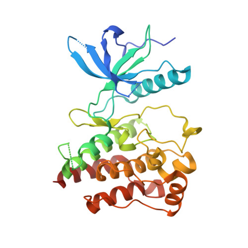
Protein-Ligand Crystal Structures Can Guide the Design of Selective Inhibitors of the FGFR Tyrosine Kinase.

Norman, R.A.Schott, A.K.Andrews, D.M.Breed, J.Foote, K.M.Garner, A.P.Ogg, D.Orme, J.P.Pink, J.H.Roberts, K.Rudge, D.A.Thomas, A.P.Leach, A.G.

(2012) J Med Chem 55: 5003-5012

  • DOI: https://doi.org/10.1021/jm3004043
  • Primary Citation Related Structures: 
    4F63, 4F64, 4F65

  • PubMed Abstract: 

    The design of compounds that selectively inhibit a single kinase is a significant challenge, particularly for compounds that bind to the ATP site. We describe here how protein-ligand crystal structure information was able both to rationalize observed selectivity and to guide the design of more selective compounds. Inhibition data from enzyme and cellular screens and the crystal structures of a range of ligands tested during the process of identifying selective inhibitors of FGFR provide a step-by-step illustration of the process. Steric effects were exploited by increasing the size of ligands in specific regions in such a way as to be tolerated in the primary target and not in other related kinases. Kinases are an excellent target class to exploit such approaches because of the conserved fold and small side chain mobility of the active form.


  • Organizational Affiliation
    • AstraZeneca Pharmaceuticals , Mereside, Alderley Park, Macclesfield, SK10 4TG, U.K.

Macromolecule Content 

  • Total Structure Weight: 71.27 kDa 
  • Atom Count: 4,319 
  • Modeled Residue Count: 549 
  • Deposited Residue Count: 618 
  • Unique protein chains: 1

Macromolecules

Find similar proteins by:|  3D Structure
Entity ID: 1
MoleculeChains  Sequence LengthOrganismDetailsImage
Fibroblast growth factor receptor 1
A, B
309Homo sapiensMutation(s): 2 
Gene Names: FGFR1BFGFRCEKFGFBRFLGFLT2HBGFR
EC: 2.7.10.1
UniProt & NIH Common Fund Data Resources
Find proteins for P11362 (Homo sapiens)
Explore P11362 
Go to UniProtKB:  P11362
PHAROS:  P11362
GTEx:  ENSG00000077782 
Entity Groups
Sequence Clusters30% Identity50% Identity70% Identity90% Identity95% Identity100% Identity
UniProt GroupP11362
Sequence Annotations
Expand
Reference Sequence

Experimental Data & Validation

Experimental Data

  • Method: X-RAY DIFFRACTION
  • Resolution: 2.55 Å
  • R-Value Free:  0.287 (Depositor), 0.285 (DCC) 
  • R-Value Work:  0.229 (Depositor), 0.236 (DCC) 
  • R-Value Observed: 0.232 (Depositor) 
Space Group: C 1 2 1
Unit Cell:
Length ( Å )Angle ( ˚ )
a = 208.306α = 90
b = 57.477β = 107.42
c = 65.452γ = 90
Software Package:
Software NamePurpose
CrystalCleardata collection
AMoREphasing
REFMACrefinement
MOSFLMdata reduction
SCALAdata scaling

Structure Validation

View Full Validation Report


Ligand Structure Quality Assessment 


Entry History 

Deposition Data

Revision History  (Full details and data files)

  • Version 1.0: 2012-06-06
    Type: Initial release
  • Version 1.1: 2012-06-27
    Changes: Database references
  • Version 1.2: 2023-09-13
    Changes: Data collection, Database references, Derived calculations, Refinement description