4F5Y | pdb_00004f5y

Crystal structure of human STING CTD complex with C-di-GMP


Experimental Data Snapshot

  • Method: X-RAY DIFFRACTION
  • Resolution: 2.40 Å
  • R-Value Free: 
    0.275 (Depositor), 0.271 (DCC) 
  • R-Value Work: 
    0.211 (Depositor), 0.207 (DCC) 
  • R-Value Observed: 
    0.215 (Depositor) 

wwPDB Validation 3D Report Full Report

Validation slider image for 4F5Y

Ligand Structure Quality Assessment 


This is version 1.2 of the entry. See complete history

Literature

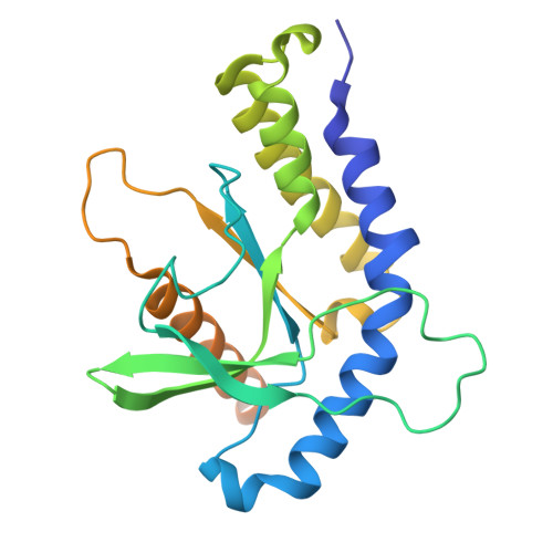
Crystal structures of STING protein reveal basis for recognition of cyclic di-GMP

Shang, G.Zhu, D.Li, N.Zhang, J.Zhu, C.Lu, D.Liu, C.Yu, Q.Zhao, Y.Xu, S.Gu, L.

(2012) Nat Struct Mol Biol 19: 725-727

  • DOI: https://doi.org/10.1038/nsmb.2332
  • Primary Citation Related Structures: 
    4F5W, 4F5Y

  • PubMed Abstract: 

    STING functions as both an adaptor protein signaling cytoplasmic double-stranded DNA and a direct immunosensor of cyclic diguanylate monophosphate (c-di-GMP). The crystal structures of the C-terminal domain of human STING (STING(CTD)) and its complex with c-di-GMP reveal how STING recognizes c-di-GMP. In response to c-di-GMP binding, two surface loops, which serve as a gate and latch of the cleft formed by the dimeric STING(CTD), undergo rearrangements to interact with the ligand.


  • Organizational Affiliation
    • State Key Laboratory of Microbial Technology, School of Life Science, Shandong University, Jinan, China.

Macromolecule Content 

  • Total Structure Weight: 55.25 kDa 
  • Atom Count: 3,292 
  • Modeled Residue Count: 386 
  • Deposited Residue Count: 478 
  • Unique protein chains: 1

Macromolecules

Find similar proteins by:|  3D Structure
Entity ID: 1
MoleculeChains  Sequence LengthOrganismDetailsImage
Transmembrane protein 173
A, B
239Homo sapiensMutation(s): 0 
Gene Names: TMEM173ERISMITASTING
UniProt & NIH Common Fund Data Resources
Find proteins for Q86WV6 (Homo sapiens)
Explore Q86WV6 
Go to UniProtKB:  Q86WV6
PHAROS:  Q86WV6
GTEx:  ENSG00000184584 
Entity Groups
Sequence Clusters30% Identity50% Identity70% Identity90% Identity95% Identity100% Identity
UniProt GroupQ86WV6
Sequence Annotations
Expand
Reference Sequence

Small Molecules

Ligands 2 Unique
IDChains Name / Formula / InChI Key2D Diagram3D Interactions
C2E

Query on C2E


Download:Ideal Coordinates CCD File
D [auth A]9,9'-[(2R,3R,3aS,5S,7aR,9R,10R,10aS,12S,14aR)-3,5,10,12-tetrahydroxy-5,12-dioxidooctahydro-2H,7H-difuro[3,2-d:3',2'-j][1,3,7,9,2,8]tetraoxadiphosphacyclododecine-2,9-diyl]bis(2-amino-1,9-dihydro-6H-purin-6-one)
C20 H24 N10 O14 P2
PKFDLKSEZWEFGL-MHARETSRSA-N
CA

Query on CA


Download:Ideal Coordinates CCD File
C [auth A],
E [auth B]
CALCIUM ION
Ca
BHPQYMZQTOCNFJ-UHFFFAOYSA-N

Experimental Data & Validation

Experimental Data

  • Method: X-RAY DIFFRACTION
  • Resolution: 2.40 Å
  • R-Value Free:  0.275 (Depositor), 0.271 (DCC) 
  • R-Value Work:  0.211 (Depositor), 0.207 (DCC) 
  • R-Value Observed: 0.215 (Depositor) 
Space Group: P 21 21 21
Unit Cell:
Length ( Å )Angle ( ˚ )
a = 73.145α = 90
b = 81.438β = 90
c = 91.347γ = 90
Software Package:
Software NamePurpose
HKL-2000data collection
PHASERphasing
PHENIXrefinement
HKL-2000data reduction
HKL-2000data scaling

Structure Validation

View Full Validation Report


Ligand Structure Quality Assessment 


Entry History 

Deposition Data

Revision History  (Full details and data files)

  • Version 1.0: 2012-06-27
    Type: Initial release
  • Version 1.1: 2013-09-04
    Changes: Database references
  • Version 1.2: 2024-03-20
    Changes: Data collection, Database references, Derived calculations, Structure summary