4F5D | pdb_00004f5d

ERIS/STING in complex with ligand


Experimental Data Snapshot

  • Method: X-RAY DIFFRACTION
  • Resolution: 3.00 Å
  • R-Value Free: 
    0.242 (Depositor), 0.236 (DCC) 
  • R-Value Work: 
    0.211 (Depositor), 0.208 (DCC) 
  • R-Value Observed: 
    0.214 (Depositor) 

wwPDB Validation 3D Report Full Report

Validation slider image for 4F5D

Ligand Structure Quality Assessment 


This is version 1.3 of the entry. See complete history

Literature

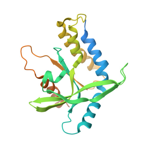
The structural basis for the sensing and binding of cyclic di-GMP by STING

Huang, Y.H.Liu, X.Y.Du, X.X.Jiang, Z.F.Su, X.D.

(2012) Nat Struct Mol Biol 19: 728-730

  • DOI: https://doi.org/10.1038/nsmb.2333
  • Primary Citation Related Structures: 
    4F5D, 4F5E

  • PubMed Abstract: 

    STING (stimulator of interferon genes) is an essential signaling adaptor that mediates cytokine production in response to microbial invasion by directly sensing bacterial secondary messengers such as the cyclic dinucleotide bis-(3'-5')-cyclic dimeric GMP (c-di-GMP). STING's structure and its binding mechanism to cyclic dinucleotides were unknown. We report here the crystal structures of the STING cytoplasmic domain and its complex with c-di-GMP, thus providing the structural basis for understanding STING function.


  • Organizational Affiliation
    • State Key Laboratory of Protein and Plant Gene Research, School of Life Sciences, Peking University, Beijing, China.

Macromolecule Content 

  • Total Structure Weight: 57.99 kDa 
  • Atom Count: 3,092 
  • Modeled Residue Count: 376 
  • Deposited Residue Count: 512 
  • Unique protein chains: 1

Macromolecules

Find similar proteins by:|  3D Structure
Entity ID: 1
MoleculeChains  Sequence LengthOrganismDetailsImage
Transmembrane protein 173
A, B
256Homo sapiensMutation(s): 1 
Gene Names: TMEM173ERISMITASTING
UniProt & NIH Common Fund Data Resources
Find proteins for Q86WV6 (Homo sapiens)
Explore Q86WV6 
Go to UniProtKB:  Q86WV6
PHAROS:  Q86WV6
GTEx:  ENSG00000184584 
Entity Groups
Sequence Clusters30% Identity50% Identity70% Identity90% Identity95% Identity100% Identity
UniProt GroupQ86WV6
Sequence Annotations
Expand
Reference Sequence

Small Molecules

Ligands 2 Unique
IDChains Name / Formula / InChI Key2D Diagram3D Interactions
C2E

Query on C2E


Download:Ideal Coordinates CCD File
C [auth A]9,9'-[(2R,3R,3aS,5S,7aR,9R,10R,10aS,12S,14aR)-3,5,10,12-tetrahydroxy-5,12-dioxidooctahydro-2H,7H-difuro[3,2-d:3',2'-j][1,3,7,9,2,8]tetraoxadiphosphacyclododecine-2,9-diyl]bis(2-amino-1,9-dihydro-6H-purin-6-one)
C20 H24 N10 O14 P2
PKFDLKSEZWEFGL-MHARETSRSA-N
MG

Query on MG


Download:Ideal Coordinates CCD File
D [auth A],
E [auth B]
MAGNESIUM ION
Mg
JLVVSXFLKOJNIY-UHFFFAOYSA-N

Experimental Data & Validation

Experimental Data

  • Method: X-RAY DIFFRACTION
  • Resolution: 3.00 Å
  • R-Value Free:  0.242 (Depositor), 0.236 (DCC) 
  • R-Value Work:  0.211 (Depositor), 0.208 (DCC) 
  • R-Value Observed: 0.214 (Depositor) 
Space Group: P 41
Unit Cell:
Length ( Å )Angle ( ˚ )
a = 111.85α = 90
b = 111.85β = 90
c = 35.02γ = 90
Software Package:
Software NamePurpose
ADSCdata collection
PHENIXmodel building
PHENIXrefinement
XDSdata reduction
XDSdata scaling
PHENIXphasing

Structure Validation

View Full Validation Report


Ligand Structure Quality Assessment 


Entry History 

Deposition Data

Revision History  (Full details and data files)

  • Version 1.0: 2012-06-27
    Type: Initial release
  • Version 1.1: 2012-11-14
    Changes: Database references
  • Version 1.2: 2017-11-15
    Changes: Refinement description
  • Version 1.3: 2024-03-20
    Changes: Data collection, Database references, Derived calculations, Refinement description, Structure summary