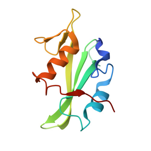
Superbinder SH2 Domains Act as Antagonists of Cell Signaling.
Kaneko, T., Huang, H., Cao, X., Li, X., Li, C., Voss, C., Sidhu, S.S., Li, S.S.(2012) Sci Signal 5: ra68-ra68
- PubMed: 23012655
- DOI: https://doi.org/10.1126/scisignal.2003021
- Primary Citation of Related Structures:
4F59, 4F5A, 4F5B - PubMed Abstract:
Protein-ligand interactions mediated by modular domains, which often play important roles in regulating cellular functions, are generally of moderate affinities. We examined the Src homology 2 (SH2) domain, a modular domain that recognizes phosphorylated tyrosine (pTyr) residues, to investigate how the binding affinity of a modular domain for its ligand influences the structure and cellular function of the protein. We used the phage display method to perform directed evolution of the pTyr-binding residues in the SH2 domain of the tyrosine kinase Fyn and identified three amino acid substitutions that critically affected binding. We generated three SH2 domain triple-point mutants that were "superbinders" with much higher affinities for pTyr-containing peptides than the natural domain. Crystallographic analysis of one of these superbinders revealed that the superbinder SH2 domain recognized the pTyr moiety in a bipartite binding mode: A hydrophobic surface encompassed the phenyl ring, and a positively charged site engaged the phosphate. When expressed in mammalian cells, the superbinder SH2 domains blocked epidermal growth factor receptor signaling and inhibited anchorage-independent cell proliferation, suggesting that pTyr superbinders might be explored for therapeutic applications and useful as biological research tools. Although the SH2 domain fold can support much higher affinity for its ligand than is observed in nature, our results suggest that natural SH2 domains are not optimized for ligand binding but for specificity and flexibility, which are likely properties important for their function in signaling and regulatory processes.
- Department of Biochemistry and Siebens-Drake Medical Research Institute, Schulich School of Medicine and Dentistry, University of Western Ontario, London, Ontario N6A 5C1, Canada.
Organizational Affiliation:


