


Crystal structure of 5-hydroxy-2'-deoxycytidine base paired with 2'-deoxyguanosine in Dickerson Drew Dodecamer
Szulik, M.W., Nocek, B., Joachimiak, A., Stone, M.P.To be published.
Experimental Data Snapshot
Starting Model: experimental
View more details
wwPDB Validation 3D Report Full Report

Entity ID: 1 | ||||
| Molecule | Chains | Length | Organism | Image |
|---|---|---|---|---|
| DNA (5'-D(*CP*GP*CP*GP*AP*AP*TP*TP*(5OC)P*GP*CP*G)-3') | 12 | synthetic construct | 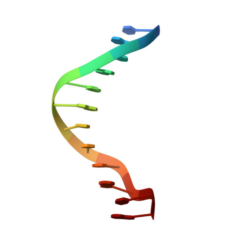 | |
Sequence AnnotationsExpand | ||||
Reference Sequence | ||||
| Ligands 1 Unique | |||||
|---|---|---|---|---|---|
| ID | Chains | Name / Formula / InChI Key | 2D Diagram | 3D Interactions | |
| MG Download:Ideal Coordinates CCD File | C [auth A] | MAGNESIUM ION Mg JLVVSXFLKOJNIY-UHFFFAOYSA-N |  | ||
| Length ( Å ) | Angle ( ˚ ) |
|---|---|
| a = 25.287 | α = 90 |
| b = 40.217 | β = 90 |
| c = 65.471 | γ = 90 |
| Software Name | Purpose |
|---|---|
| SBC-Collect | data collection |
| MOLREP | phasing |
| REFMAC | refinement |
| HKL-3000 | data reduction |
| HKL-3000 | data scaling |


