4F3T | pdb_00004f3t

Human Argonaute-2 - miR-20a complex


Experimental Data Snapshot

  • Method: X-RAY DIFFRACTION
  • Resolution: 2.25 Å
  • R-Value Free: 
    0.254 (Depositor), 0.247 (DCC) 
  • R-Value Work: 
    0.207 (Depositor), 0.202 (DCC) 
  • R-Value Observed: 
    0.208 (Depositor) 

wwPDB Validation 3D Report Full Report

Validation slider image for 4F3T

This is version 1.3 of the entry. See complete history

Literature

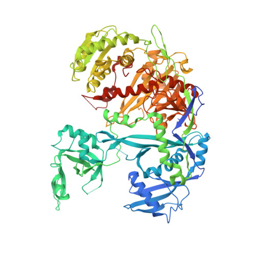
The Structure of Human Argonaute-2 in Complex with miR-20a.

Elkayam, E.Kuhn, C.D.Tocilj, A.Haase, A.D.Greene, E.M.Hannon, G.J.Joshua-Tor, L.

(2012) Cell 150: 100-110

  • DOI: https://doi.org/10.1016/j.cell.2012.05.017
  • Primary Citation Related Structures: 
    4F3T

  • PubMed Abstract: 

    Argonaute proteins lie at the heart of the RNA-induced silencing complex (RISC), wherein they use small RNA guides to recognize targets. Initial insight into the architecture of Argonautes came from studies of prokaryotic proteins, revealing a crescent-shaped base made up of the amino-terminal, PAZ, middle, and PIWI domains. The recently reported crystal structure of human Argonaute-2 (hAgo2), the "slicer" in RNA interference, in complex with a mixed population of RNAs derived from insect cells provides insight into the architecture of a eukaryotic Argonaute protein with defined biochemical and biological functions. Here, we report the structure of human Ago2 bound to a physiologically relevant microRNA, microRNA-20a, at 2.2 Å resolution. The miRNA is anchored at both ends by the Mid and PAZ domains and makes several kinks and turns along the binding groove. Interestingly, miRNA binding confers remarkable stability on hAgo2, locking this otherwise flexible enzyme into a stable conformation.


  • Organizational Affiliation
    • W. M. Keck Structural Biology Laboratory, 1 Bungtown Road, Cold Spring Harbor, NY 11724, USA.

Macromolecule Content 

  • Total Structure Weight: 104.12 kDa 
  • Atom Count: 6,884 
  • Modeled Residue Count: 815 
  • Deposited Residue Count: 881 
  • Unique protein chains: 1
  • Unique nucleic acid chains: 1

Macromolecules


Find similar proteins by:|  3D Structure
Entity ID: 1
MoleculeChains  Sequence LengthOrganismDetailsImage
Protein argonaute-2861Homo sapiensMutation(s): 0 
Gene Names: AGO2Argonaute2eIF-2C 2eIF2C 2EIF2C2Eukaryotic translation initiation factor 2C 2hAgo2Human Argonaute-2PAZ Piwi domain protein
EC: 3.1.26
UniProt & NIH Common Fund Data Resources
Find proteins for Q9UKV8 (Homo sapiens)
Explore Q9UKV8 
Go to UniProtKB:  Q9UKV8
PHAROS:  Q9UKV8
GTEx:  ENSG00000123908 
Entity Groups
Sequence Clusters30% Identity50% Identity70% Identity90% Identity95% Identity100% Identity
UniProt GroupQ9UKV8
Sequence Annotations
Expand
Reference Sequence
Find similar nucleic acids by:  Sequence
Entity ID: 2
MoleculeChains LengthOrganismImage
RNA (5'-R(P*UP*AP*AP*AP*GP*UP*GP*CP*UP*UP*AP*UP*AP*GP*UP*G*CP*AP*GP*G)-3')B [auth R]20Homo sapiens
Sequence Annotations
Expand
Reference Sequence

Experimental Data & Validation

Experimental Data

  • Method: X-RAY DIFFRACTION
  • Resolution: 2.25 Å
  • R-Value Free:  0.254 (Depositor), 0.247 (DCC) 
  • R-Value Work:  0.207 (Depositor), 0.202 (DCC) 
  • R-Value Observed: 0.208 (Depositor) 
Space Group: P 1 21 1
Unit Cell:
Length ( Å )Angle ( ˚ )
a = 63.427α = 90
b = 107.651β = 106.69
c = 68.718γ = 90
Software Package:
Software NamePurpose
DENZOdata reduction
SCALEPACKdata scaling
SHARPphasing
SOLOMONphasing
PHENIXrefinement
PDB_EXTRACTdata extraction
HKL-2000data collection

Structure Validation

View Full Validation Report


Entry History 

Deposition Data

Revision History  (Full details and data files)

  • Version 1.0: 2012-05-30
    Type: Initial release
  • Version 1.1: 2012-07-25
    Changes: Database references
  • Version 1.2: 2017-11-15
    Changes: Advisory, Refinement description
  • Version 1.3: 2024-02-28
    Changes: Advisory, Data collection, Database references, Derived calculations