4F3D | pdb_00004f3d

Structure of RPE65: P65 crystal form grown in Fos-Choline-10


Experimental Data Snapshot

  • Method: X-RAY DIFFRACTION
  • Resolution: 2.50 Å
  • R-Value Free: 
    0.219 (Depositor), 0.221 (DCC) 
  • R-Value Work: 
    0.193 (Depositor), 0.194 (DCC) 
  • R-Value Observed: 
    0.194 (Depositor) 

Starting Model: experimental
View more details

wwPDB Validation 3D Report Full Report

Validation slider image for 4F3D

This is version 1.2 of the entry. See complete history

Literature

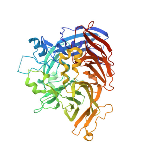
Structure of RPE65 isomerase in a lipidic matrix reveals roles for phospholipids and iron in catalysis.

Kiser, P.D.Farquhar, E.R.Shi, W.Sui, X.Chance, M.R.Palczewski, K.

(2012) Proc Natl Acad Sci U S A 109: E2747-E2756

  • DOI: https://doi.org/10.1073/pnas.1212025109
  • Primary Citation Related Structures: 
    4F2Z, 4F30, 4F3A, 4F3D

  • PubMed Abstract: 

    RPE65 is a key metalloenzyme responsible for maintaining visual function in vertebrates. Despite extensive research on this membrane-bound retinoid isomerase, fundamental questions regarding its enzymology remain unanswered. Here, we report the crystal structure of RPE65 in a membrane-like environment. These crystals, obtained from enzymatically active, nondelipidated protein, displayed an unusual packing arrangement wherein RPE65 is embedded in a lipid-detergent sheet. Structural differences between delipidated and nondelipidated RPE65 uncovered key residues involved in substrate uptake and processing. Complementary iron K-edge X-ray absorption spectroscopy data established that RPE65 as isolated contained a divalent iron center and demonstrated the presence of a tightly bound ligand consistent with a coordinated carboxylate group. These results support the hypothesis that the Lewis acidity of iron could be used to promote ester dissociation and generation of a carbocation intermediate required for retinoid isomerization.


  • Organizational Affiliation
    • Department of Pharmacology, School of Medicine, Case Western Reserve University, Cleveland, OH 44106, USA. pdk7@case.edu

Macromolecule Content 

  • Total Structure Weight: 122.46 kDa 
  • Atom Count: 8,421 
  • Modeled Residue Count: 1,026 
  • Deposited Residue Count: 1,066 
  • Unique protein chains: 1

Macromolecules

Find similar proteins by:|  3D Structure
Entity ID: 1
MoleculeChains  Sequence LengthOrganismDetailsImage
Retinoid isomerohydrolase
A, B
533Bos taurusMutation(s): 0 
EC: 3.1.1.64 (PDB Primary Data), 5.3.3.22 (UniProt)
Membrane Entity: Yes 
UniProt
Find proteins for Q28175 (Bos taurus)
Explore Q28175 
Go to UniProtKB:  Q28175
Entity Groups
Sequence Clusters30% Identity50% Identity70% Identity90% Identity95% Identity100% Identity
UniProt GroupQ28175
Sequence Annotations
Expand
Reference Sequence

Experimental Data & Validation

Experimental Data

  • Method: X-RAY DIFFRACTION
  • Resolution: 2.50 Å
  • R-Value Free:  0.219 (Depositor), 0.221 (DCC) 
  • R-Value Work:  0.193 (Depositor), 0.194 (DCC) 
  • R-Value Observed: 0.194 (Depositor) 
Space Group: P 65
Unit Cell:
Length ( Å )Angle ( ˚ )
a = 175.258α = 90
b = 175.258β = 90
c = 86.449γ = 120
Software Package:
Software NamePurpose
XDSdata scaling
REFMACrefinement
XDSdata reduction

Structure Validation

View Full Validation Report


Entry History 

Deposition Data

Revision History  (Full details and data files)

  • Version 1.0: 2012-10-03
    Type: Initial release
  • Version 1.1: 2014-07-02
    Changes: Database references
  • Version 1.2: 2023-09-13
    Changes: Data collection, Database references, Derived calculations, Refinement description