4F2A | pdb_00004f2a

Crystal structure of cholestryl esters transfer protein in complex with inhibitors


Experimental Data Snapshot

  • Method: X-RAY DIFFRACTION
  • Resolution: 3.11 Å
  • R-Value Free: 
    0.239 (Depositor), 0.251 (DCC) 
  • R-Value Work: 
    0.183 (Depositor), 0.203 (DCC) 
  • R-Value Observed: 
    0.185 (Depositor) 

wwPDB Validation 3D Report Full Report

Validation slider image for 4F2A

Ligand Structure Quality Assessment 


This is version 2.1 of the entry. See complete history

Literature

Crystal structures of cholesteryl ester transfer protein in complex with inhibitors.

Liu, S.Mistry, A.Reynolds, J.M.Lloyd, D.B.Griffor, M.C.Perry, D.A.Ruggeri, R.B.Clark, R.W.Qiu, X.

(2012) J Biological Chem 287: 37321-37329

  • DOI: https://doi.org/10.1074/jbc.M112.380063
  • Primary Citation Related Structures: 
    4EWS, 4F2A

  • PubMed Abstract: 

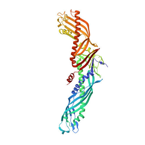
    Human plasma cholesteryl ester transfer protein (CETP) transports cholesteryl ester from the antiatherogenic high-density lipoproteins (HDL) to the proatherogenic low-density and very low-density lipoproteins (LDL and VLDL). Inhibition of CETP has been shown to raise human plasma HDL cholesterol (HDL-C) levels and is potentially a novel approach for the prevention of cardiovascular diseases. Here, we report the crystal structures of CETP in complex with torcetrapib, a CETP inhibitor that has been tested in phase 3 clinical trials, and compound 2, an analog from a structurally distinct inhibitor series. In both crystal structures, the inhibitors are buried deeply within the protein, shifting the bound cholesteryl ester in the N-terminal pocket of the long hydrophobic tunnel and displacing the phospholipid from that pocket. The lipids in the C-terminal pocket of the hydrophobic tunnel remain unchanged. The inhibitors are positioned near the narrowing neck of the hydrophobic tunnel of CETP and thus block the connection between the N- and C-terminal pockets. These structures illuminate the unusual inhibition mechanism of these compounds and support the tunnel mechanism for neutral lipid transfer by CETP. These highly lipophilic inhibitors bind mainly through extensive hydrophobic interactions with the protein and the shifted cholesteryl ester molecule. However, polar residues, such as Ser-230 and His-232, are also found in the inhibitor binding site. An enhanced understanding of the inhibitor binding site may provide opportunities to design novel CETP inhibitors possessing more drug-like physical properties, distinct modes of action, or alternative pharmacological profiles.


  • Organizational Affiliation
    • Department of Structural Biology & Biophysics, Pfizer Groton Laboratories, Groton, Connecticut 06340, USA. shenping.liu@pfizer.com

Macromolecule Content 

  • Total Structure Weight: 56.67 kDa 
  • Atom Count: 3,946 
  • Modeled Residue Count: 472 
  • Deposited Residue Count: 476 
  • Unique protein chains: 1

Macromolecules

Find similar proteins by:|  3D Structure
Entity ID: 1
MoleculeChains  Sequence LengthOrganismDetailsImage
Cholesteryl ester transfer protein476Homo sapiensMutation(s): 6 
Gene Names: CETP
UniProt & NIH Common Fund Data Resources
Find proteins for P11597 (Homo sapiens)
Explore P11597 
Go to UniProtKB:  P11597
PHAROS:  P11597
GTEx:  ENSG00000087237 
Entity Groups
Sequence Clusters30% Identity50% Identity70% Identity90% Identity95% Identity100% Identity
UniProt GroupP11597
Glycosylation
Glycosylation Sites: 1Go to GlyGen: P11597-1
Sequence Annotations
Expand
Reference Sequence

Oligosaccharides

Help  
Entity ID: 2
MoleculeChains Length2D Diagram GlycosylationD Interactions
alpha-D-mannopyranose-(1-4)-2-acetamido-2-deoxy-beta-D-glucopyranose-(1-4)-[alpha-L-fucopyranose-(1-6)]2-acetamido-2-deoxy-beta-D-glucopyranose
B
4N-Glycosylation
Glycosylation Resources
GlyTouCan: G47477HI
GlyCosmos: G47477HI
GlyGen: G47477HI

Small Molecules

Ligands 5 Unique
IDChains Name / Formula / InChI Key2D Diagram3D Interactions
PCW

Query on PCW


Download:Ideal Coordinates CCD File
E [auth A]1,2-DIOLEOYL-SN-GLYCERO-3-PHOSPHOCHOLINE
C44 H85 N O8 P
SNKAWJBJQDLSFF-NVKMUCNASA-O
2OB

Query on 2OB


Download:Ideal Coordinates CCD File
C [auth A],
D [auth A]
CHOLESTERYL OLEATE
C45 H78 O2
RJECHNNFRHZQKU-RMUVNZEASA-N
0SF

Query on 0SF


Download:Ideal Coordinates CCD File
H [auth A](2R)-3-{[4-(4-chloro-3-ethylphenoxy)pyrimidin-2-yl][3-(1,1,2,2-tetrafluoroethoxy)benzyl]amino}-1,1,1-trifluoropropan-2-ol
C24 H21 Cl F7 N3 O3
BTIIJGFMCVLCET-LJQANCHMSA-N
PGE

Query on PGE


Download:Ideal Coordinates CCD File
F [auth A]TRIETHYLENE GLYCOL
C6 H14 O4
ZIBGPFATKBEMQZ-UHFFFAOYSA-N
CL

Query on CL


Download:Ideal Coordinates CCD File
G [auth A]CHLORIDE ION
Cl
VEXZGXHMUGYJMC-UHFFFAOYSA-M

Experimental Data & Validation

Experimental Data

  • Method: X-RAY DIFFRACTION
  • Resolution: 3.11 Å
  • R-Value Free:  0.239 (Depositor), 0.251 (DCC) 
  • R-Value Work:  0.183 (Depositor), 0.203 (DCC) 
  • R-Value Observed: 0.185 (Depositor) 
Space Group: P 21 21 21
Unit Cell:
Length ( Å )Angle ( ˚ )
a = 68.772α = 90
b = 69.915β = 90
c = 187.145γ = 90
Software Package:
Software NamePurpose
BUSTERrefinement

Structure Validation

View Full Validation Report


Ligand Structure Quality Assessment 


Entry History 

Deposition Data

Revision History  (Full details and data files)

  • Version 1.0: 2012-09-05
    Type: Initial release
  • Version 1.1: 2012-09-26
    Changes: Database references
  • Version 1.2: 2012-11-14
    Changes: Database references
  • Version 2.0: 2020-07-29
    Type: Remediation
    Reason: Carbohydrate remediation
    Changes: Advisory, Atomic model, Data collection, Database references, Derived calculations, Structure summary
  • Version 2.1: 2024-11-20
    Changes: Data collection, Database references, Structure summary