4F25 | pdb_00004f25

Crystal structure of the second RRM domain of human PABPC1 at pH 6.0


Experimental Data Snapshot

  • Method: X-RAY DIFFRACTION
  • Resolution: 1.90 Å
  • R-Value Free: 
    0.269 (Depositor), 0.263 (DCC) 
  • R-Value Work: 
    0.217 (Depositor), 0.216 (DCC) 
  • R-Value Observed: 
    0.219 (Depositor) 

Starting Model: experimental
View more details

wwPDB Validation 3D Report Full Report

Validation slider image for 4F25

This is version 1.2 of the entry. See complete history

Literature

Interdomain Allostery Promotes Assembly of the Poly(A) mRNA Complex with PABP and eIF4G.

Safaee, N.Kozlov, G.Noronha, A.M.Xie, J.Wilds, C.J.Gehring, K.

(2012) Mol Cell 48: 375-386

  • DOI: https://doi.org/10.1016/j.molcel.2012.09.001
  • Primary Citation Related Structures: 
    4F02, 4F25, 4F26

  • PubMed Abstract: 

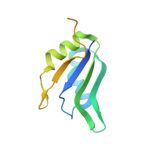
    Many RNA-binding proteins contain multiple single-strand nucleic acid-binding domains and assemble into large multiprotein messenger ribonucleic acid protein (mRNP) complexes. The mechanisms underlying the self-assembly of these complexes are largely unknown. In eukaryotes, the association of the translation factors polyadenylate-binding protein-1 (PABP) and eIF4G is essential for high-level expression of polyadenylated mRNAs. Here, we report the crystal structure of the ternary complex poly(A)(11)·PABP(1-190)·eIF4G(178-203) at 2.0 Å resolution. Our NMR and crystallographic data show that eIF4G interacts with the RRM2 domain of PABP. Analysis of the interaction by small-angle X-ray scattering, isothermal titration calorimetry, and electromobility shift assays reveals that this interaction is allosterically regulated by poly(A) binding to PABP. Furthermore, we have confirmed the importance of poly(A) for the endogenous PABP and eIF4G interaction in immunoprecipitation experiments using HeLa cell extracts. Our findings reveal interdomain allostery as a mechanism for cooperative assembly of RNP complexes.


  • Organizational Affiliation
    • Department of Biochemistry, McGill University, Montréal, QC H3G 0B1, Canada.

Macromolecule Content 

  • Total Structure Weight: 12.68 kDa 
  • Atom Count: 700 
  • Modeled Residue Count: 81 
  • Deposited Residue Count: 115 
  • Unique protein chains: 1

Macromolecules

Find similar proteins by:|  3D Structure
Entity ID: 1
MoleculeChains  Sequence LengthOrganismDetailsImage
Polyadenylate-binding protein 1115Homo sapiensMutation(s): 0 
Gene Names: PABPC1PAB1PABP1PABPC2
UniProt & NIH Common Fund Data Resources
Find proteins for P11940 (Homo sapiens)
Explore P11940 
Go to UniProtKB:  P11940
PHAROS:  P11940
GTEx:  ENSG00000070756 
Entity Groups
Sequence Clusters30% Identity50% Identity70% Identity90% Identity95% Identity100% Identity
UniProt GroupP11940
Sequence Annotations
Expand
Reference Sequence

Experimental Data & Validation

Experimental Data

  • Method: X-RAY DIFFRACTION
  • Resolution: 1.90 Å
  • R-Value Free:  0.269 (Depositor), 0.263 (DCC) 
  • R-Value Work:  0.217 (Depositor), 0.216 (DCC) 
  • R-Value Observed: 0.219 (Depositor) 
Space Group: P 31 2 1
Unit Cell:
Length ( Å )Angle ( ˚ )
a = 50.653α = 90
b = 50.653β = 90
c = 63.447γ = 120
Software Package:
Software NamePurpose
ADSCdata collection
PHASERphasing
REFMACrefinement
HKL-2000data reduction
HKL-2000data scaling

Structure Validation

View Full Validation Report


Entry History 

Deposition Data

Revision History  (Full details and data files)

  • Version 1.0: 2012-08-08
    Type: Initial release
  • Version 1.1: 2013-01-02
    Changes: Database references
  • Version 1.2: 2023-09-13
    Changes: Data collection, Database references, Refinement description