4EXG | pdb_00004exg

Design and synthesis of potent hydroxyethylamine (hea) bace-1 inhibitors


Experimental Data Snapshot

  • Method: X-RAY DIFFRACTION
  • Resolution: 1.80 Å
  • R-Value Free: 
    0.227 (Depositor), 0.233 (DCC) 
  • R-Value Work: 
    0.178 (Depositor), 0.178 (DCC) 
  • R-Value Observed: 
    0.181 (Depositor) 

wwPDB Validation 3D Report Full Report

Validation slider image for 4EXG

Ligand Structure Quality Assessment 


This is version 1.1 of the entry. See complete history

Literature

Design and synthesis of potent hydroxyethylamine (HEA) BACE-1 inhibitors carrying prime side 4,5,6,7-tetrahydrobenzazole and 4,5,6,7-tetrahydropyridinoazole templates.

Sund, C.Belda, O.Borkakoti, N.Lindberg, J.Derbyshire, D.Vrang, L.Hamelink, E.Hgren, C.Woestenenk, E.Wikstrom, K.Eneroth, A.Lindstrom, E.Kalayanov, G.

(2012) Bioorg Med Chem Lett 22: 6721-6727

  • DOI: https://doi.org/10.1016/j.bmcl.2012.08.097
  • Primary Citation Related Structures: 
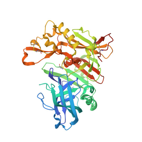
    4EWO, 4EXG

  • PubMed Abstract: 

    A set of low molecular weight compounds containing a hydroxyethylamine (HEA) core structure with different prime side alkyl substituted 4,5,6,7-tetrahydrobenzazoles and one 4,5,6,7-tetrahydropyridinoazole was synthesized. Striking differences were observed on potencies in the BACE-1 enzymatic and cellular assays depending on the nature of the heteroatoms in the bicyclic ring, from the low active compound 4 to inhibitor 6, displaying BACE-1 IC(50) values of 44 nM (enzyme assay) and 65 nM (cell-based assay).


  • Organizational Affiliation
    • Medivir AB, Lunastigen 7, SE-14144 Huddinge, Sweden.

Macromolecule Content 

  • Total Structure Weight: 43.48 kDa 
  • Atom Count: 3,504 
  • Modeled Residue Count: 377 
  • Deposited Residue Count: 386 
  • Unique protein chains: 1

Macromolecules

Find similar proteins by:|  3D Structure
Entity ID: 1
MoleculeChains  Sequence LengthOrganismDetailsImage
Beta-secretase 1386Homo sapiensMutation(s): 0 
Gene Names: BACE1BACEKIAA1149
EC: 3.4.23.46
UniProt & NIH Common Fund Data Resources
Find proteins for P56817 (Homo sapiens)
Explore P56817 
Go to UniProtKB:  P56817
PHAROS:  P56817
GTEx:  ENSG00000186318 
Entity Groups
Sequence Clusters30% Identity50% Identity70% Identity90% Identity95% Identity100% Identity
UniProt GroupP56817
Sequence Annotations
Expand
Reference Sequence

Small Molecules

Ligands 1 Unique
IDChains Name / Formula / InChI Key2D Diagram3D Interactions
916

Query on 916


Download:Ideal Coordinates CCD File
B [auth A]N-[(2S,3R)-4-{[(4S)-6-(2,2-dimethylpropyl)-2,2-dimethyl-3,4-dihydro-2H-thieno[2,3-b]pyran-4-yl]amino}-3-hydroxy-1-phenylbutan-2-yl]acetamide
C26 H38 N2 O3 S
YLIBYDKWVPKMKX-RJGXRXQPSA-N

Experimental Data & Validation

Experimental Data

  • Method: X-RAY DIFFRACTION
  • Resolution: 1.80 Å
  • R-Value Free:  0.227 (Depositor), 0.233 (DCC) 
  • R-Value Work:  0.178 (Depositor), 0.178 (DCC) 
  • R-Value Observed: 0.181 (Depositor) 
Space Group: P 21 21 21
Unit Cell:
Length ( Å )Angle ( ˚ )
a = 48.223α = 90
b = 76.371β = 90
c = 104.377γ = 90
Software Package:
Software NamePurpose
REFMACrefinement
PDB_EXTRACTdata extraction
DNAdata collection
MOSFLMdata reduction
SCALEPACKdata scaling
PHASERphasing

Structure Validation

View Full Validation Report


Ligand Structure Quality Assessment 


Entry History 

Deposition Data

Revision History  (Full details and data files)

  • Version 1.0: 2012-10-31
    Type: Initial release
  • Version 1.1: 2024-11-20
    Changes: Data collection, Database references, Derived calculations, Structure summary