4EWT

The crystal structure of a putative aminohydrolase from methicillin resistant Staphylococcus aureus


Experimental Data Snapshot

  • Method: X-RAY DIFFRACTION
  • Resolution: 2.10 Å
  • R-Value Free: 0.229 
  • R-Value Work: 0.199 
  • R-Value Observed: 0.200 

wwPDB Validation   3D Report Full Report


Ligand Structure Quality Assessment 


This is version 1.1 of the entry. See complete history


Literature

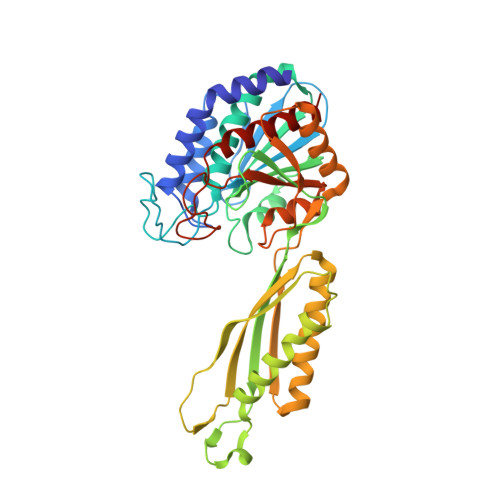
Structure of an amidohydrolase, SACOL0085, from methicillin-resistant Staphylococcus aureus COL

Girish, T.S.Vivek, B.Colaco, M.Misquith, S.Gopal, B.

(2013) Acta Crystallogr Sect F Struct Biol Cryst Commun 69: 103-108

  • DOI: https://doi.org/10.1107/S1744309112049822
  • Primary Citation of Related Structures:  
    4EWT

  • PubMed Abstract: 

    Staphylococcus aureus is an opportunistic pathogen that rapidly acquires resistance to frontline antibiotics. The characterization of novel protein targets from this bacterium is thus an important step towards future therapeutic strategies. Here, the crystal structure of an amidohydrolase, SACOL0085, from S. aureus COL is described. SACOL0085 is a member of the M20D family of peptidases. Unlike other M20D peptidases, which are either monomers or dimers, SACOL0085 adopts a butterfly-shaped homotetrameric arrangement with extensive intersubunit interactions. Each subunit of SACOL0085 contains two Mn(2+) ions at the active site. A conserved cysteine residue at the active site distinguishes M20D peptidases from other M20 family members. This cysteine, Cys103, serves as bidentate ligand coordinating both Mn(2+) ions in SACOL0085.


  • Organizational Affiliation

    Molecular Biophysics Unit, Indian Institute of Science, Bangalore 560 012, India.


Macromolecules
Find similar proteins by:  (by identity cutoff)  |  3D Structure
Entity ID: 1
MoleculeChains Sequence LengthOrganismDetailsImage
Peptidase, M20/M25/M40 familyA,
B [auth C],
C [auth D],
D [auth B]
392Staphylococcus aureus subsp. aureus COLMutation(s): 0 
Gene Names: SACOL0085
EC: 3.5.1.14
UniProt
Find proteins for A0A0H2WZV8 (Staphylococcus aureus (strain COL))
Explore A0A0H2WZV8 
Go to UniProtKB:  A0A0H2WZV8
Entity Groups  
Sequence Clusters30% Identity50% Identity70% Identity90% Identity95% Identity100% Identity
UniProt GroupA0A0H2WZV8
Sequence Annotations
Expand
  • Reference Sequence
Small Molecules
Ligands 3 Unique
IDChains Name / Formula / InChI Key2D Diagram3D Interactions
PE7
Query on PE7

Download Ideal Coordinates CCD File 
L [auth D],
O [auth B],
P [auth B]
1-DEOXY-1-THIO-HEPTAETHYLENE GLYCOL
C14 H30 O7 S
ACMBXVJDKVNCGH-UHFFFAOYSA-N
PEG
Query on PEG

Download Ideal Coordinates CCD File 
G [auth A]DI(HYDROXYETHYL)ETHER
C4 H10 O3
MTHSVFCYNBDYFN-UHFFFAOYSA-N
MN
Query on MN

Download Ideal Coordinates CCD File 
E [auth A]
F [auth A]
H [auth C]
I [auth C]
J [auth D]
E [auth A],
F [auth A],
H [auth C],
I [auth C],
J [auth D],
K [auth D],
M [auth B],
N [auth B]
MANGANESE (II) ION
Mn
WAEMQWOKJMHJLA-UHFFFAOYSA-N
Experimental Data & Validation

Experimental Data

  • Method: X-RAY DIFFRACTION
  • Resolution: 2.10 Å
  • R-Value Free: 0.229 
  • R-Value Work: 0.199 
  • R-Value Observed: 0.200 
  • Space Group: P 1
Unit Cell:
Length ( Å )Angle ( ˚ )
a = 44.62α = 115.4
b = 120.11β = 94.64
c = 132.41γ = 96.55
Software Package:
Software NamePurpose
MAR345dtbdata collection
PHASERphasing
REFMACrefinement
MOSFLMdata reduction
SCALAdata scaling

Structure Validation

View Full Validation Report


Ligand Structure Quality Assessment 


Entry History 

Deposition Data

Revision History  (Full details and data files)

  • Version 1.0: 2013-02-20
    Type: Initial release
  • Version 1.1: 2023-11-08
    Changes: Data collection, Database references, Derived calculations, Refinement description