


Crystal structure of a strand-swapped dimer of Mouse Leukocyte-associated immunoglobulin-like receptor 1 Extra Cellular Domain
Sampathkumar, P., Ramagopal, U.A., Bonanno, J., Fiser, A., Zencheck, W., Nathenson, S.G., Almo, S.C.To be published.
Experimental Data Snapshot
Starting Model: other
View more details
wwPDB Validation 3D Report Full Report

Macromolecule Content
Entity ID: 1 | |||||
|---|---|---|---|---|---|
| Molecule | Chains | Sequence Length | Organism | Details | Image |
| Leukocyte-associated immunoglobulin-like receptor 1 | 118 | Mus musculus | Mutation(s): 0 Gene Names: Lair1 | 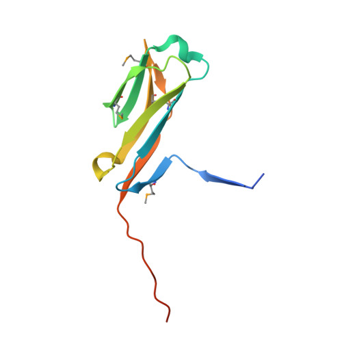 | |
UniProt | |||||
Entity Groups | |||||
| Sequence Clusters | 30% Identity50% Identity70% Identity90% Identity95% Identity100% Identity | ||||
| UniProt Group | Q8BG84 | ||||
Sequence AnnotationsExpand | |||||
Reference Sequence | |||||
| Ligands 1 Unique | |||||
|---|---|---|---|---|---|
| ID | Chains | Name / Formula / InChI Key | 2D Diagram | 3D Interactions | |
| EDO Download:Ideal Coordinates CCD File | E [auth A] F [auth A] G [auth B] H [auth B] I [auth B] | 1,2-ETHANEDIOL C2 H6 O2 LYCAIKOWRPUZTN-UHFFFAOYSA-N |  | ||
| Modified Residues 1 Unique | |||||
|---|---|---|---|---|---|
| ID | Chains | Type | Formula | 2D Diagram | Parent |
| MSE Query on MSE | A, B, C, D | L-PEPTIDE LINKING | C5 H11 N O2 Se |  | MET |
| Length ( Å ) | Angle ( ˚ ) |
|---|---|
| a = 175.588 | α = 90 |
| b = 57.427 | β = 105.15 |
| c = 47.795 | γ = 90 |
| Software Name | Purpose |
|---|---|
| REFMAC | refinement |
| PDB_EXTRACT | data extraction |
| ADSC | data collection |
| HKL-3000 | data reduction |
| HKL-3000 | data scaling |
| PHENIX | phasing |


