4ET0 | pdb_00004et0

Crystal structure of circularly permuted human asparaginase-like protein 1


Experimental Data Snapshot

  • Method: X-RAY DIFFRACTION
  • Resolution: 3.30 Å
  • R-Value Free: 
    0.249 (Depositor), 0.301 (DCC) 
  • R-Value Work: 
    0.214 (Depositor), 0.260 (DCC) 
  • R-Value Observed: 
    0.216 (Depositor) 

wwPDB Validation 3D Report Full Report

Validation slider image for 4ET0

This is version 1.2 of the entry. See complete history

Literature

Uncoupling Intramolecular Processing and Substrate Hydrolysis in the N-Terminal Nucleophile Hydrolase hASRGL1 by Circular Permutation.

Li, W.Cantor, J.R.Yogesha, S.D.Yang, S.Chantranupong, L.Liu, J.Q.Agnello, G.Georgiou, G.Stone, E.M.Zhang, Y.

(2012) ACS Chem Biol 7: 1840-1847

  • DOI: https://doi.org/10.1021/cb300232n
  • Primary Citation Related Structures: 
    3TKJ, 4ET0

  • PubMed Abstract: 

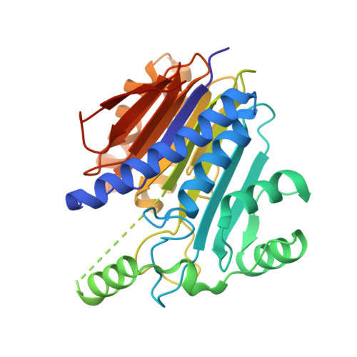
    The human asparaginase-like protein 1 (hASRGL1) catalyzes the hydrolysis of l-asparagine and isoaspartyl-dipeptides. As an N-terminal nucleophile (Ntn) hydrolase superfamily member, the active form of hASRGL1 is generated by an intramolecular cleavage step with Thr168 as the catalytic residue. However, in vitro, autoprocessing is incomplete (~50%), fettering the biophysical characterization of hASRGL1. We circumvented this obstacle by constructing a circularly permuted hASRGL1 that uncoupled the autoprocessing reaction, allowing us to kinetically and structurally characterize this enzyme and the precursor-like hASRGL1-Thr168Ala variant. Crystallographic and biochemical evidence suggest an activation mechanism where a torsional restraint on the Thr168 side chain helps drive the intramolecular processing reaction. Cleavage and formation of the active site releases the torsional restriction on Thr168, which is facilitated by a small conserved Gly-rich loop near the active site that allows the conformational changes necessary for activation.


  • Organizational Affiliation
    • Department of Chemistry and Biochemistry, University of Texas, Austin, Texas 78712, United States.

Macromolecule Content 

  • Total Structure Weight: 67.58 kDa 
  • Atom Count: 4,304 
  • Modeled Residue Count: 593 
  • Deposited Residue Count: 654 
  • Unique protein chains: 1

Macromolecules

Find similar proteins by:|  3D Structure
Entity ID: 1
MoleculeChains  Sequence LengthOrganismDetailsImage
L-asparaginase
A, B
327Homo sapiensMutation(s): 0 
Gene Names: ASRGL1ALPCRASH
EC: 3.5.1.1 (PDB Primary Data), 3.4.19.5 (UniProt)
UniProt & NIH Common Fund Data Resources
Find proteins for Q7L266 (Homo sapiens)
Explore Q7L266 
Go to UniProtKB:  Q7L266
PHAROS:  Q7L266
GTEx:  ENSG00000162174 
Entity Groups
Sequence Clusters30% Identity50% Identity70% Identity90% Identity95% Identity100% Identity
UniProt GroupQ7L266
Sequence Annotations
Expand
Reference Sequence

Experimental Data & Validation

Experimental Data

  • Method: X-RAY DIFFRACTION
  • Resolution: 3.30 Å
  • R-Value Free:  0.249 (Depositor), 0.301 (DCC) 
  • R-Value Work:  0.214 (Depositor), 0.260 (DCC) 
  • R-Value Observed: 0.216 (Depositor) 
Space Group: P 65 2 2
Unit Cell:
Length ( Å )Angle ( ˚ )
a = 108.638α = 90
b = 108.638β = 90
c = 275.144γ = 120
Software Package:
Software NamePurpose
HKL-2000data collection
PHASERphasing
BUSTERrefinement
HKL-2000data reduction
HKL-2000data scaling

Structure Validation

View Full Validation Report


Entry History 

Deposition Data

Revision History  (Full details and data files)

  • Version 1.0: 2012-09-26
    Type: Initial release
  • Version 1.1: 2012-12-12
    Changes: Database references
  • Version 1.2: 2024-02-28
    Changes: Data collection, Database references, Derived calculations