4ERS

A Molecular Basis for Negative Regulation of the Glucagon Receptor


Experimental Data Snapshot

  • Method: X-RAY DIFFRACTION
  • Resolution: 2.64 Å
  • R-Value Free: 0.279 
  • R-Value Work: 0.231 
  • R-Value Observed: 0.234 

wwPDB Validation   3D Report Full Report


Ligand Structure Quality Assessment 


This is version 1.3 of the entry. See complete history


Literature

Molecular basis for negative regulation of the glucagon receptor.

Koth, C.M.Murray, J.M.Mukund, S.Madjidi, A.Minn, A.Clarke, H.J.Wong, T.Chiang, V.Luis, E.Estevez, A.Rondon, J.Zhang, Y.Hotzel, I.Allan, B.B.

(2012) Proc Natl Acad Sci U S A 109: 14393-14398

  • DOI: https://doi.org/10.1073/pnas.1206734109
  • Primary Citation of Related Structures:  
    4ERS

  • PubMed Abstract: 

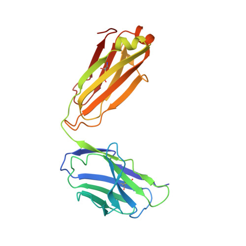
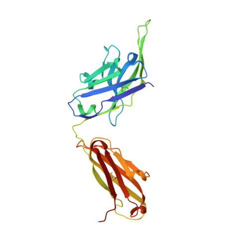
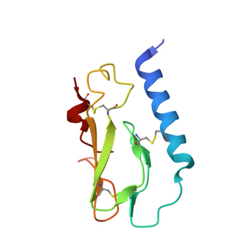
    Members of the class B family of G protein-coupled receptors (GPCRs) bind peptide hormones and have causal roles in many diseases, ranging from diabetes and osteoporosis to anxiety. Although peptide, small-molecule, and antibody inhibitors of these GPCRs have been identified, structure-based descriptions of receptor antagonism are scarce. Here we report the mechanisms of glucagon receptor inhibition by blocking antibodies targeting the receptor's extracellular domain (ECD). These studies uncovered a role for the ECD as an intrinsic negative regulator of receptor activity. The crystal structure of the ECD in complex with the Fab fragment of one antibody, mAb1, reveals that this antibody inhibits glucagon receptor by occluding a surface extending across the entire hormone-binding cleft. A second antibody, mAb23, blocks glucagon binding and inhibits basal receptor activity, indicating that it is an inverse agonist and that the ECD can negatively regulate receptor activity independent of ligand binding. Biochemical analyses of receptor mutants in the context of a high-resolution ECD structure show that this previously unrecognized inhibitory activity of the ECD involves an interaction with the third extracellular loop of the receptor and suggest that glucagon-mediated structural changes in the ECD accompany receptor activation. These studies have implications for the design of drugs to treat class B GPCR-related diseases, including the potential for developing novel allosteric regulators that target the ECDs of these receptors.


  • Organizational Affiliation

    Department of Structural Biology, Molecular Biology, Antibody Engineering, Protein Chemistry, and Early Discovery Biochemistry, Genentech Inc, South San Francisco, CA 94080, USA. koth.christopher@gene.com


Macromolecules
Find similar proteins by:  (by identity cutoff)  |  3D Structure
Entity ID: 1
MoleculeChains Sequence LengthOrganismDetailsImage
Fab light chainA [auth L]214Homo sapiensMutation(s): 0 
Entity Groups  
Sequence Clusters30% Identity50% Identity70% Identity90% Identity95% Identity100% Identity
Sequence Annotations
Expand
  • Reference Sequence
Find similar proteins by:  (by identity cutoff)  |  3D Structure
Entity ID: 2
MoleculeChains Sequence LengthOrganismDetailsImage
Fab heavy chainB [auth H]231Homo sapiensMutation(s): 0 
Entity Groups  
Sequence Clusters30% Identity50% Identity70% Identity90% Identity95% Identity100% Identity
Sequence Annotations
Expand
  • Reference Sequence
Find similar proteins by:  (by identity cutoff)  |  3D Structure
Entity ID: 3
MoleculeChains Sequence LengthOrganismDetailsImage
Glucagon receptorC [auth A]96Homo sapiensMutation(s): 0 
Gene Names: GCGR
UniProt & NIH Common Fund Data Resources
Find proteins for P47871 (Homo sapiens)
Explore P47871 
Go to UniProtKB:  P47871
PHAROS:  P47871
GTEx:  ENSG00000215644 
Entity Groups  
Sequence Clusters30% Identity50% Identity70% Identity90% Identity95% Identity100% Identity
UniProt GroupP47871
Sequence Annotations
Expand
  • Reference Sequence
Small Molecules
Ligands 1 Unique
IDChains Name / Formula / InChI Key2D Diagram3D Interactions
NAG
Query on NAG

Download Ideal Coordinates CCD File 
D [auth A],
E [auth A]
2-acetamido-2-deoxy-beta-D-glucopyranose
C8 H15 N O6
OVRNDRQMDRJTHS-FMDGEEDCSA-N
Experimental Data & Validation

Experimental Data

  • Method: X-RAY DIFFRACTION
  • Resolution: 2.64 Å
  • R-Value Free: 0.279 
  • R-Value Work: 0.231 
  • R-Value Observed: 0.234 
  • Space Group: P 1 21 1
Unit Cell:
Length ( Å )Angle ( ˚ )
a = 46.41α = 90
b = 62.25β = 93.91
c = 104.94γ = 90
Software Package:
Software NamePurpose
SCALAdata scaling
PHASERphasing
PHENIXrefinement
PDB_EXTRACTdata extraction
XSCALEdata scaling

Structure Validation

View Full Validation Report


Ligand Structure Quality Assessment 


Entry History 

Deposition Data

Revision History  (Full details and data files)

  • Version 1.0: 2012-08-29
    Type: Initial release
  • Version 1.1: 2012-10-17
    Changes: Database references
  • Version 1.2: 2012-12-05
    Changes: Derived calculations
  • Version 1.3: 2020-07-29
    Type: Remediation
    Reason: Carbohydrate remediation
    Changes: Advisory, Data collection, Derived calculations, Structure summary