4ENE | pdb_00004ene

Structure of the N- and C-terminal trimmed ClC-ec1 Cl-/H+ antiporter and Fab Complex


Experimental Data Snapshot

  • Method: X-RAY DIFFRACTION
  • Resolution: 2.40 Å
  • R-Value Free: 
    0.278 (Depositor), 0.277 (DCC) 
  • R-Value Work: 
    0.239 (Depositor), 0.236 (DCC) 
  • R-Value Observed: 
    0.241 (Depositor) 

wwPDB Validation 3D Report Full Report

Validation slider image for 4ENE

Ligand Structure Quality Assessment 


This is version 2.1 of the entry. See complete history

Literature

Intracellular proton access in a cl(-)/h(+) antiporter.

Lim, H.H.Shane, T.Miller, C.

(2012) PLoS Biol 10: e1001441-e1001441

  • DOI: https://doi.org/10.1371/journal.pbio.1001441
  • Primary Citation Related Structures: 
    4ENE

  • PubMed Abstract: 

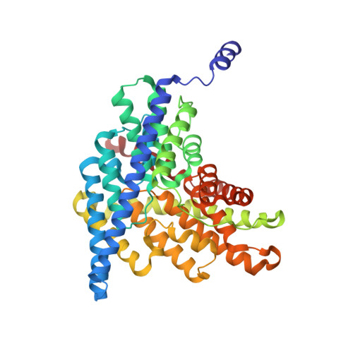
    Chloride-transporting membrane proteins of the CLC family appear in two distinct mechanistic flavors: H(+)-gated Cl(-) channels and Cl(-)/H(+) antiporters. Transmembrane H(+) movement is an essential feature of both types of CLC. X-ray crystal structures of CLC antiporters show the Cl(-) ion pathway through these proteins, but the H(+) pathway is known only inferentially by two conserved glutamate residues that act as way-stations for H(+) in its path through the protein. The extracellular-facing H(+) transfer glutamate becomes directly exposed to aqueous solution during the transport cycle, but the intracellular glutamate E203, Glu(in), is buried within the protein. Two regions, denoted "polar" and "interfacial," at the intracellular surface of the bacterial antiporter CLC-ec1 are examined here as possible pathways by which intracellular aqueous protons gain access to Glu(in). Mutations at multiple residues of the polar region have little effect on antiport rates. In contrast, mutation of E202, a conserved glutamate at the protein-water boundary of the interfacial region, leads to severe slowing of the Cl(-)/H(+) antiport rate. An X-ray crystal structure of E202Y, the most strongly inhibited of these substitutions, shows an aqueous portal leading to Glu(in) physically blocked by cross-subunit interactions; moreover, this mutation has only minimal effect on a monomeric CLC variant, which necessarily lacks such interactions. The several lines of experiments presented argue that E202 acts as a water-organizer that creates a proton conduit connecting intracellular solvent with Glu(in).


  • Organizational Affiliation
    • Department of Biochemistry, Howard Hughes Medical Institute, Brandeis University, Waltham, Massachusetts, USA.

Macromolecule Content 

  • Total Structure Weight: 193.16 kDa 
  • Atom Count: 13,691 
  • Modeled Residue Count: 1,751 
  • Deposited Residue Count: 1,758 
  • Unique protein chains: 3

Macromolecules

Find similar proteins by:|  3D Structure
Entity ID: 1
MoleculeChains  Sequence LengthOrganismDetailsImage
H(+)/Cl(-) exchange transporter ClcA
A, B
446Escherichia coli K-12Mutation(s): 0 
Gene Names: clcAeriCyadQb0155JW5012
Membrane Entity: Yes 
UniProt
Find proteins for P37019 (Escherichia coli (strain K12))
Explore P37019 
Go to UniProtKB:  P37019
Entity Groups
Sequence Clusters30% Identity50% Identity70% Identity90% Identity95% Identity100% Identity
UniProt GroupP37019
Sequence Annotations
Expand
Reference Sequence
Find similar proteins by:|  3D Structure
Entity ID: 2
MoleculeChains  Sequence LengthOrganismDetailsImage
heavy chain of Fab fragment
C, E
222Mus musculusMutation(s): 0 
Find similar proteins by:|  3D Structure
Entity ID: 3
MoleculeChains  Sequence LengthOrganismDetailsImage
light chain of Fab fragment
D, F
211Mus musculusMutation(s): 0 

Oligosaccharides

Help  
Entity ID: 4
MoleculeChains Length2D Diagram GlycosylationD Interactions
alpha-D-glucopyranose-(1-4)-alpha-D-glucopyranose
G, H, I
2N/A
Glycosylation Resources
GlyTouCan: G07411ON
GlyCosmos: G07411ON

Small Molecules

Ligands 2 Unique
IDChains Name / Formula / InChI Key2D Diagram3D Interactions
DMU

Query on DMU


Download:Ideal Coordinates CCD File
L [auth A]
M [auth A]
N [auth A]
O [auth A]
S [auth B]
L [auth A],
M [auth A],
N [auth A],
O [auth A],
S [auth B],
T [auth B]
DECYL-BETA-D-MALTOPYRANOSIDE
C22 H42 O11
WOQQAWHSKSSAGF-WXFJLFHKSA-N
CL

Query on CL


Download:Ideal Coordinates CCD File
J [auth A],
K [auth A],
P [auth B],
Q [auth B],
R [auth B]
CHLORIDE ION
Cl
VEXZGXHMUGYJMC-UHFFFAOYSA-M

Biologically Interesting Molecules (External Reference) 

1 Unique

Experimental Data & Validation

Experimental Data

  • Method: X-RAY DIFFRACTION
  • Resolution: 2.40 Å
  • R-Value Free:  0.278 (Depositor), 0.277 (DCC) 
  • R-Value Work:  0.239 (Depositor), 0.236 (DCC) 
  • R-Value Observed: 0.241 (Depositor) 
Space Group: C 1 2 1
Unit Cell:
Length ( Å )Angle ( ˚ )
a = 232.26α = 90
b = 97.52β = 132.87
c = 173.96γ = 90
Software Package:
Software NamePurpose
ADSCdata collection
PHASERphasing
PHENIXrefinement
MOSFLMdata reduction
SCALAdata scaling

Structure Validation

View Full Validation Report


Ligand Structure Quality Assessment 


Entry History 

Deposition Data

Revision History  (Full details and data files)

  • Version 1.0: 2012-12-26
    Type: Initial release
  • Version 1.1: 2018-01-24
    Changes: Structure summary
  • Version 2.0: 2020-07-29
    Type: Remediation
    Reason: Carbohydrate remediation
    Changes: Atomic model, Data collection, Database references, Derived calculations, Non-polymer description, Structure summary
  • Version 2.1: 2024-10-16
    Changes: Data collection, Database references, Structure summary