


Crystal structure of the ferric binding protein A
Wang, Q., Liu, X.Q., Wang, X.Q.To be published.
Experimental Data Snapshot
wwPDB Validation 3D Report Full Report

Macromolecule Content
Entity ID: 1 | |||||
|---|---|---|---|---|---|
| Molecule | Chains | Sequence Length | Organism | Details | Image |
| Iron ABC transporter, periplasmic iron-binding protein | 336 | Thermus thermophilus HB8 | Mutation(s): 0 Gene Names: TTHA1628 | 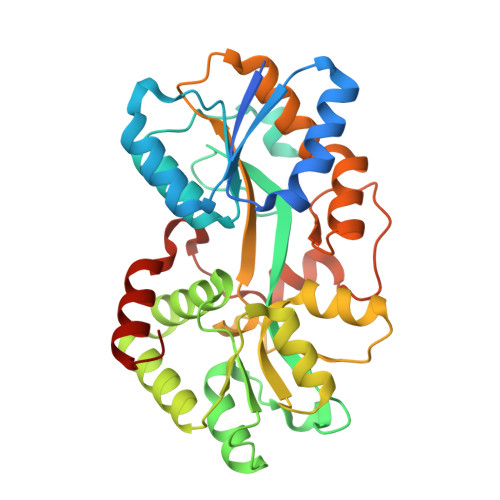 | |
UniProt | |||||
Entity Groups | |||||
| Sequence Clusters | 30% Identity50% Identity70% Identity90% Identity95% Identity100% Identity | ||||
| UniProt Group | Q5SHV2 | ||||
Sequence AnnotationsExpand | |||||
Reference Sequence | |||||
| Length ( Å ) | Angle ( ˚ ) |
|---|---|
| a = 42.124 | α = 90 |
| b = 139.349 | β = 90 |
| c = 326.337 | γ = 90 |
| Software Name | Purpose |
|---|---|
| HKL-2000 | data collection |
| PHASER | phasing |
| PHENIX | refinement |
| HKL-2000 | data reduction |
| HKL-2000 | data scaling |


