4ELJ | pdb_00004elj

Crystal structure of the inactive retinoblastoma protein phosphorylated at T373


Experimental Data Snapshot

  • Method: X-RAY DIFFRACTION
  • Resolution: 2.70 Å
  • R-Value Free: 
    0.266 (Depositor), 0.259 (DCC) 
  • R-Value Work: 
    0.212 (Depositor), 0.207 (DCC) 
  • R-Value Observed: 
    0.216 (Depositor) 

wwPDB Validation 3D Report Full Report

Validation slider image for 4ELJ

This is version 1.3 of the entry. See complete history

Literature

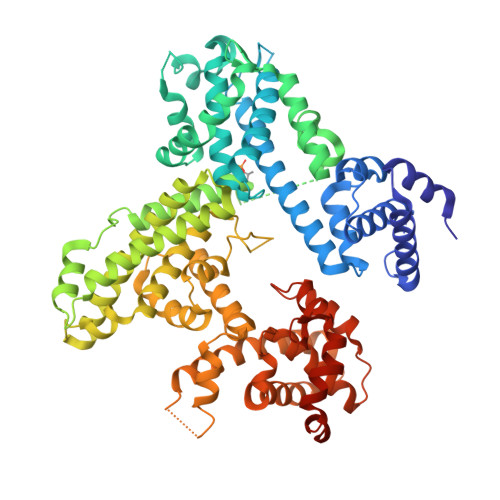
Structures of inactive retinoblastoma protein reveal multiple mechanisms for cell cycle control.

Burke, J.R.Hura, G.L.Rubin, S.M.

(2012) Genes Dev 26: 1156-1166

  • DOI: https://doi.org/10.1101/gad.189837.112
  • Primary Citation Related Structures: 
    4ELJ, 4ELL

  • PubMed Abstract: 

    Cyclin-dependent kinase (Cdk) phosphorylation of the Retinoblastoma protein (Rb) drives cell proliferation through inhibition of Rb complexes with E2F transcription factors and other regulatory proteins. We present the first structures of phosphorylated Rb that reveal the mechanism of its inactivation. S608 phosphorylation orders a flexible "pocket" domain loop such that it mimics and directly blocks E2F transactivation domain (E2F(TD)) binding. T373 phosphorylation induces a global conformational change that associates the pocket and N-terminal domains (RbN). This first multidomain Rb structure demonstrates a novel role for RbN in allosterically inhibiting the E2F(TD)-pocket association and protein binding to the pocket "LxCxE" site. Together, these structures detail the regulatory mechanism for a canonical growth-repressive complex and provide a novel example of how multisite Cdk phosphorylation induces diverse structural changes to influence cell cycle signaling.


  • Organizational Affiliation
    • Department of Chemistry and Biochemistry, University of California at Santa Cruz, Santa Cruz, California 95064, USA.

Macromolecule Content 

  • Total Structure Weight: 76.37 kDa 
  • Atom Count: 4,908 
  • Modeled Residue Count: 590 
  • Deposited Residue Count: 656 
  • Unique protein chains: 1

Macromolecules

Find similar proteins by:|  3D Structure
Entity ID: 1
MoleculeChains  Sequence LengthOrganismDetailsImage
Retinoblastoma-associated protein656Homo sapiensMutation(s): 0 
Gene Names: RB1
UniProt & NIH Common Fund Data Resources
Find proteins for P06400 (Homo sapiens)
Explore P06400 
Go to UniProtKB:  P06400
PHAROS:  P06400
GTEx:  ENSG00000139687 
Entity Groups
Sequence Clusters30% Identity50% Identity70% Identity90% Identity95% Identity100% Identity
UniProt GroupP06400
Sequence Annotations
Expand
Reference Sequence

Small Molecules

Modified Residues  1 Unique
IDChains TypeFormula2D DiagramParent
TPO
Query on TPO
A
L-PEPTIDE LINKINGC4 H10 N O6 PTHR

Experimental Data & Validation

Experimental Data

  • Method: X-RAY DIFFRACTION
  • Resolution: 2.70 Å
  • R-Value Free:  0.266 (Depositor), 0.259 (DCC) 
  • R-Value Work:  0.212 (Depositor), 0.207 (DCC) 
  • R-Value Observed: 0.216 (Depositor) 
Space Group: P 21 21 21
Unit Cell:
Length ( Å )Angle ( ˚ )
a = 51.62α = 90
b = 129.51β = 90
c = 135.04γ = 90
Software Package:
Software NamePurpose
JBluIce-EPICSdata collection
PHASERphasing
PHENIXrefinement
MOSFLMdata reduction
SCALAdata scaling

Structure Validation

View Full Validation Report


Entry History 

Deposition Data

Revision History  (Full details and data files)

  • Version 1.0: 2012-05-23
    Type: Initial release
  • Version 1.1: 2012-08-15
    Changes: Database references
  • Version 1.2: 2017-08-23
    Changes: Refinement description, Source and taxonomy
  • Version 1.3: 2024-11-06
    Changes: Data collection, Database references, Derived calculations, Structure summary