4EIQ | pdb_00004eiq

Chromopyrrolic acid-soaked RebC-10x with bound 7-carboxy-K252c


Experimental Data Snapshot

  • Method: X-RAY DIFFRACTION
  • Resolution: 2.76 Å
  • R-Value Free: 
    0.255 (Depositor), 0.255 (DCC) 
  • R-Value Work: 
    0.206 (Depositor), 0.206 (DCC) 

wwPDB Validation 3D Report Full Report

Validation slider image for 4EIQ

Ligand Structure Quality Assessment 


This is version 1.1 of the entry. See complete history

Literature

An Unusual Role for a Mobile Flavin in StaC-like Indolocarbazole Biosynthetic Enzymes.

Goldman, P.J.Ryan, K.S.Hamill, M.J.Howard-Jones, A.R.Walsh, C.T.Elliott, S.J.Drennan, C.L.

(2012) Chem Biol 19: 855-865

  • DOI: https://doi.org/10.1016/j.chembiol.2012.05.016
  • Primary Citation Related Structures: 
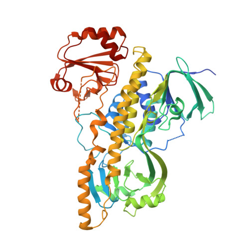
    4EIP, 4EIQ

  • PubMed Abstract: 

    The indolocarbazole biosynthetic enzymes StaC, InkE, RebC, and AtmC mediate the degree of oxidation of chromopyrrolic acid on route to the natural products staurosporine, K252a, rebeccamycin, and AT2433-A1, respectively. Here, we show that StaC and InkE, which mediate a net 4-electron oxidation, bind FAD with a micromolar K(d), whereas RebC and AtmC, which mediate a net 8-electron oxidation, bind FAD with a nanomolar K(d) while displaying the same FAD redox properties. We further create RebC-10x, a RebC protein with ten StaC-like amino acid substitutions outside of previously characterized FAD-binding motifs and the complementary StaC-10x. We find that these mutations mediate both FAD affinity and product specificity, with RebC-10x displaying higher StaC activity than StaC itself. X-ray structures of this StaC catalyst identify the substrate of StaC as 7-carboxy-K252c and suggest a unique mechanism for this FAD-dependent enzyme.


  • Organizational Affiliation
    • Department of Chemistry, Massachusetts Institute of Technology, Cambridge, MA 02139, USA.

Macromolecule Content 

  • Total Structure Weight: 120.07 kDa 
  • Atom Count: 7,928 
  • Modeled Residue Count: 1,025 
  • Deposited Residue Count: 1,098 
  • Unique protein chains: 1

Macromolecules

Find similar proteins by:|  3D Structure
Entity ID: 1
MoleculeChains  Sequence LengthOrganismDetailsImage
Putative FAD-monooxygenase
A, B
549Lentzea aerocolonigenesMutation(s): 10 
Gene Names: rbmDrebC
UniProt
Find proteins for Q8KI25 (Lentzea aerocolonigenes)
Explore Q8KI25 
Go to UniProtKB:  Q8KI25
Entity Groups
Sequence Clusters30% Identity50% Identity70% Identity90% Identity95% Identity100% Identity
UniProt GroupQ8KI25
Sequence Annotations
Expand
Reference Sequence

Small Molecules

Ligands 1 Unique
IDChains Name / Formula / InChI Key2D Diagram3D Interactions
KCT

Query on KCT


Download:Ideal Coordinates CCD File
C [auth A],
D [auth B]
(5S)-7-oxo-6,7,12,13-tetrahydro-5H-indolo[2,3-a]pyrrolo[3,4-c]carbazole-5-carboxylic acid
C21 H13 N3 O3
LYSTWLDKIFYXOG-IBGZPJMESA-N

Experimental Data & Validation

Experimental Data

  • Method: X-RAY DIFFRACTION
  • Resolution: 2.76 Å
  • R-Value Free:  0.255 (Depositor), 0.255 (DCC) 
  • R-Value Work:  0.206 (Depositor), 0.206 (DCC) 
Space Group: P 1 21 1
Unit Cell:
Length ( Å )Angle ( ˚ )
a = 63.184α = 90
b = 77.703β = 98.78
c = 123.145γ = 90
Software Package:
Software NamePurpose
APEXdata collection
CNSrefinement
HKL-2000data reduction
HKL-2000data scaling
CNSphasing

Structure Validation

View Full Validation Report


Ligand Structure Quality Assessment 


Entry History 

Deposition Data

Revision History  (Full details and data files)

  • Version 1.0: 2012-08-08
    Type: Initial release
  • Version 1.1: 2024-02-28
    Changes: Data collection, Database references, Derived calculations