4EHF | pdb_00004ehf

Allosteric Modulation of Caspase-3 through Mutagenesis


Experimental Data Snapshot

  • Method: X-RAY DIFFRACTION
  • Resolution: 1.66 Å
  • R-Value Free: 
    0.185 (Depositor), 0.183 (DCC) 
  • R-Value Work: 
    0.162 (Depositor), 0.160 (DCC) 
  • R-Value Observed: 
    0.163 (Depositor) 

wwPDB Validation 3D Report Full Report

Validation slider image for 4EHF

This is version 1.4 of the entry. See complete history

Literature

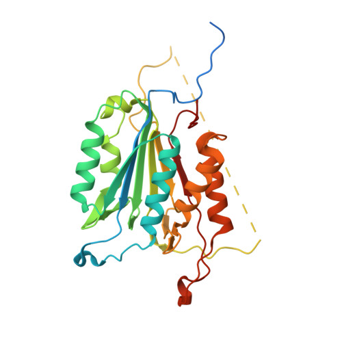
Allosteric modulation of caspase 3 through mutagenesis.

Walters, J.Schipper, J.L.Swartz, P.Mattos, C.Clark, A.C.

(2012) Biosci Rep 32: 401-411

  • DOI: https://doi.org/10.1042/BSR20120037
  • Primary Citation Related Structures: 
    4EHA, 4EHD, 4EHF, 4EHH, 4EHK, 4EHL, 4EHN

  • PubMed Abstract: 

    A mutation in the allosteric site of the caspase 3 dimer interface of Val266 to histidine abolishes activity of the enzyme, and models predict that the mutation mimics the action of small molecule allosteric inhibitors by preventing formation of the active site. Mutations were coupled to His266 at two sites in the interface, E124A and Y197C. We present results from X-ray crystallography, enzymatic activity and molecular dynamics simulations for seven proteins, consisting of single, double and triple mutants. The results demonstrate that considering allosteric inhibition of caspase 3 as a shift between discrete 'off-state' or 'on-state' conformations is insufficient. Although His266 is accommodated in the interface, the structural defects are propagated to the active site through a helix on the protein surface. A more comprehensive view of allosteric regulation of caspase 3 requires the representation of an ensemble of inactive states and shows that subtle structural changes lead to the population of the inactive ensemble.


  • Organizational Affiliation
    • Department of Molecular and Structural Biochemistry, North Carolina State University, Raleigh, NC 27695, USA.

Macromolecule Content 

  • Total Structure Weight: 32.17 kDa 
  • Atom Count: 2,269 
  • Modeled Residue Count: 248 
  • Deposited Residue Count: 283 
  • Unique protein chains: 2

Macromolecules

Find similar proteins by:|  3D Structure
Entity ID: 1
MoleculeChains  Sequence LengthOrganismDetailsImage
Caspase-3277Homo sapiensMutation(s): 2 
Gene Names: CASP3CPP32
EC: 3.4.22.56
UniProt & NIH Common Fund Data Resources
Find proteins for P42574 (Homo sapiens)
Explore P42574 
Go to UniProtKB:  P42574
PHAROS:  P42574
GTEx:  ENSG00000164305 
Entity Groups
Sequence Clusters30% Identity50% Identity70% Identity90% Identity95% Identity100% Identity
UniProt GroupP42574
Sequence Annotations
Expand
Reference Sequence
Find similar proteins by:  Sequence   |   3D Structure  
Entity ID: 2
MoleculeChains  Sequence LengthOrganismDetailsImage
ACE-ASP-GLU-VAL-ASP-CHLOROMETHYLKETONE INHIBITOR6synthetic constructMutation(s): 0 

Biologically Interesting Molecules (External Reference) 

1 Unique

Experimental Data & Validation

Experimental Data

  • Method: X-RAY DIFFRACTION
  • Resolution: 1.66 Å
  • R-Value Free:  0.185 (Depositor), 0.183 (DCC) 
  • R-Value Work:  0.162 (Depositor), 0.160 (DCC) 
  • R-Value Observed: 0.163 (Depositor) 
Space Group: I 2 2 2
Unit Cell:
Length ( Å )Angle ( ˚ )
a = 68.547α = 90
b = 84.596β = 90
c = 96.48γ = 90
Software Package:
Software NamePurpose
MAR345data collection
SERGUIdata collection
PHENIXmodel building
PHENIXrefinement
HKL-2000data reduction
HKL-2000data scaling
PHENIXphasing

Structure Validation

View Full Validation Report


Entry History 

Deposition Data

Revision History  (Full details and data files)

  • Version 1.0: 2012-06-06
    Type: Initial release
  • Version 1.1: 2012-07-18
    Changes: Database references
  • Version 1.2: 2012-12-12
    Changes: Other
  • Version 1.3: 2017-11-15
    Changes: Advisory, Refinement description, Source and taxonomy
  • Version 1.4: 2024-10-16
    Changes: Advisory, Data collection, Database references, Derived calculations, Structure summary