


Crystal structure of D-alanine-D-alanine ligase B from Burkholderia pseudomallei
Edwards, T.E., Fairman, J.W., Abendroth, J., Seattle Structural Genomics Center for Infectious Disease (SSGCID)To be published.
Experimental Data Snapshot
Starting Model: experimental
View more details
wwPDB Validation 3D Report Full Report

Entity ID: 1 | |||||
|---|---|---|---|---|---|
| Molecule | Chains | Sequence Length | Organism | Details | Image |
| D-alanine--D-alanine ligase | 316 | Burkholderia pseudomallei 1710b | Mutation(s): 0 Gene Names: BURPS1710b_3542, ddl EC: 6.3.2.4 | 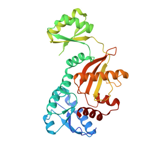 | |
UniProt | |||||
Entity Groups | |||||
| Sequence Clusters | 30% Identity50% Identity70% Identity90% Identity95% Identity100% Identity | ||||
| UniProt Group | Q3JNE1 | ||||
Sequence AnnotationsExpand | |||||
Reference Sequence | |||||
| Length ( Å ) | Angle ( ˚ ) |
|---|---|
| a = 45.389 | α = 84.47 |
| b = 66.092 | β = 80.5 |
| c = 98.558 | γ = 83.59 |
| Software Name | Purpose |
|---|---|
| DENZO | data reduction |
| SCALEPACK | data scaling |
| PHASER | phasing |
| REFMAC | refinement |
| PDB_EXTRACT | data extraction |


