4EGC

Crystal Structure of MBP-fused Human Six1 Bound to Human Eya2 Eya Domain


Experimental Data Snapshot

  • Method: X-RAY DIFFRACTION
  • Resolution: 1.99 Å
  • R-Value Free: 0.224 
  • R-Value Work: 0.184 
  • R-Value Observed: 0.186 

wwPDB Validation   3D Report Full Report


This is version 2.0 of the entry. See complete history


Literature

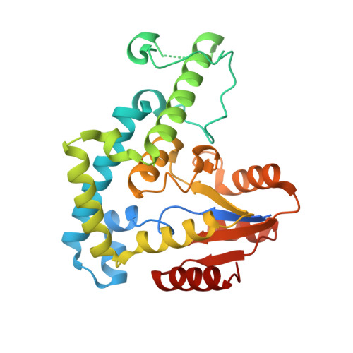
Structure-function analyses of the human SIX1-EYA2 complex reveal insights into metastasis and BOR syndrome.

Patrick, A.N.Cabrera, J.H.Smith, A.L.Chen, X.S.Ford, H.L.Zhao, R.

(2013) Nat Struct Mol Biol 20: 447-453

  • DOI: https://doi.org/10.1038/nsmb.2505
  • Primary Citation of Related Structures:  
    4EGC

  • PubMed Abstract: 
  • SIX1 interacts with EYA to form a bipartite transcription factor essential for mammalian development. Loss of function of this complex causes branchio-oto-renal (BOR) syndrome, whereas re-expression of SIX1 or EYA promotes metastasis. Here we describe the 2 ...

    SIX1 interacts with EYA to form a bipartite transcription factor essential for mammalian development. Loss of function of this complex causes branchio-oto-renal (BOR) syndrome, whereas re-expression of SIX1 or EYA promotes metastasis. Here we describe the 2.0-Å structure of SIX1 bound to EYA2, which suggests a new DNA-binding mechanism for SIX1 and provides a rationale for the effect of BOR syndrome mutations. The structure also reveals that SIX1 uses predominantly a single helix to interact with EYA. Substitution of a single amino acid in this helix is sufficient to disrupt SIX1-EYA interaction, SIX1-mediated epithelial-mesenchymal transition and metastasis in mouse models. Given that SIX1 and EYA are overexpressed in many tumor types, our data indicate that targeting the SIX1-EYA complex may be a potent approach to inhibit tumor progression in multiple cancer types.


    Organizational Affiliation

    Department of Obstetrics and Gynecology, University of Colorado Anschutz Medical Campus, Aurora, Colorado, USA.


Macromolecules
Find similar proteins by:  (by identity cutoff)  |  3D Structure
Entity ID: 1
MoleculeChainsSequence LengthOrganismDetailsImage
Maltose-binding periplasmic protein, Homeobox protein SIX1 chimeraA559Escherichia coliHomo sapiens
This entity is chimeric
Mutation(s): 5 
Gene Names: b4034JW3994malEMBPSIX1
UniProt & NIH Common Fund Data Resources
Find proteins for P0AEX9 (Escherichia coli (strain K12))
Explore P0AEX9 
Go to UniProtKB:  P0AEX9
Find proteins for Q15475 (Homo sapiens)
Explore Q15475 
Go to UniProtKB:  Q15475
PHAROS:  Q15475
Entity Groups  
Sequence Clusters30% Identity50% Identity70% Identity90% Identity95% Identity100% Identity
UniProt GroupsQ15475P0AEX9
Protein Feature View
Expand
  • Reference Sequence
Find similar proteins by:  (by identity cutoff)  |  3D Structure
Entity ID: 2
MoleculeChainsSequence LengthOrganismDetailsImage
Eyes absent homolog 2B294Homo sapiensMutation(s): 0 
Gene Names: EAB1EYA2
EC: 3.1.3.48
UniProt & NIH Common Fund Data Resources
Find proteins for O00167 (Homo sapiens)
Explore O00167 
Go to UniProtKB:  O00167
PHAROS:  O00167
Entity Groups  
Sequence Clusters30% Identity50% Identity70% Identity90% Identity95% Identity100% Identity
UniProt GroupO00167
Protein Feature View
Expand
  • Reference Sequence
Oligosaccharides

Help

Entity ID: 3
MoleculeChainsChain Length2D DiagramGlycosylation3D Interactions
alpha-D-glucopyranose-(1-4)-alpha-D-glucopyranoseC 2N/A Oligosaccharides Interaction
Glycosylation Resources
GlyTouCan:  G07411ON
GlyCosmos:  G07411ON
Small Molecules
Ligands 1 Unique
IDChainsName / Formula / InChI Key2D Diagram3D Interactions
MG
Query on MG

Download Ideal Coordinates CCD File 
D [auth B]MAGNESIUM ION
Mg
JLVVSXFLKOJNIY-UHFFFAOYSA-N
 Ligand Interaction
Biologically Interesting Molecules (External Reference) 1 Unique
Entity ID: 3
IDChainsNameType/Class2D Diagram3D Interactions
PRD_900001
Query on PRD_900001
Calpha-maltoseOligosaccharide / Nutrient Ligand Interaction
Experimental Data & Validation

Experimental Data

  • Method: X-RAY DIFFRACTION
  • Resolution: 1.99 Å
  • R-Value Free: 0.224 
  • R-Value Work: 0.184 
  • R-Value Observed: 0.186 
  • Space Group: P 21 21 2
Unit Cell:
Length ( Å )Angle ( ˚ )
a = 123.207α = 90
b = 150.18β = 90
c = 53.921γ = 90
Software Package:
Software NamePurpose
DENZOdata reduction
SCALEPACKdata scaling
PHENIXrefinement
PDB_EXTRACTdata extraction
HKL-3000data collection
MLPHAREphasing

Structure Validation

View Full Validation Report


Entry History 

Deposition Data

Revision History  (Full details and data files)

  • Version 1.0: 2013-02-27
    Type: Initial release
  • Version 1.1: 2013-03-20
    Changes: Database references
  • Version 1.2: 2013-04-17
    Changes: Database references
  • Version 1.3: 2017-07-26
    Changes: Refinement description, Source and taxonomy
  • Version 2.0: 2020-07-29
    Type: Remediation
    Reason: Carbohydrate remediation
    Changes: Atomic model, Data collection, Database references, Derived calculations, Non-polymer description, Structure summary