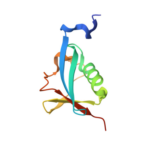
Crystal structure of the ubiquitin-like domain of human TBK1.
Li, J., Li, J., Miyahira, A., Sun, J., Liu, Y., Cheng, G., Liang, H.(2012) Protein Cell 3: 383-391
- PubMed: 22610919
- DOI: https://doi.org/10.1007/s13238-012-2929-1
- Primary Citation of Related Structures:
4EFO - PubMed Abstract:
TANK-binding kinase 1 (TBK1) is an important enzyme in the regulation of cellular antiviral effects. TBK1 regulates the activity of the interferon regulatory factors IRF3 and IRF7, thereby playing a key role in type I interferon (IFN) signaling pathways. The structure of TBK1 consists of an N-terminal kinase domain, a middle ubiquitin-like domain (ULD), and a C-terminal elongated helical domain. It has been reported that the ULD of TBK1 regulates kinase activity, playing an important role in signaling and mediating interactions with other molecules in the IFN pathway. In this study, we present the crystal structure of the ULD of human TBK1 and identify several conserved residues by multiple sequence alignment. We found that a hydrophobic patch in TBK1, containing residues Leu316, Ile353, and Val382, corresponding to the "Ile44 hydrophobic patch" observed in ubiquitin, was conserved in TBK1, IκB kinase epsilon (IKKɛ/IKKi), IκB kinase alpha (IKKα), and IκB kinase beta (IKKβ). In comparison with the structure of the IKKβ ULD domain of Xenopus laevis, we speculate that the Ile44 hydrophobic patch of TBK1 is present in an intramolecular binding surface between ULD and the C-terminal elongated helices. The varying surface charge distributions in the ULD domains of IKK and IKK-related kinases may be relevant to their specificity for specific partners.
- State Key Laboratory of Biomacromolecules, Institute of Biophysics, Chinese Academy of Sciences, Beijing 100101, China.
Organizational Affiliation:


