4EFN | pdb_00004efn

Crystal structure of H-Ras Q61L in complex with GppNHp (state 1)


Experimental Data Snapshot

  • Method: X-RAY DIFFRACTION
  • Resolution: 2.30 Å
  • R-Value Free: 
    0.237 (Depositor), 0.233 (DCC) 
  • R-Value Work: 
    0.183 (Depositor), 0.181 (DCC) 
  • R-Value Observed: 
    0.185 (Depositor) 

Starting Model: experimental
View more details

wwPDB Validation 3D Report Full Report

Validation slider image for 4EFN

Ligand Structure Quality Assessment 


This is version 1.2 of the entry. See complete history

Literature

Crystal structures of the state 1 conformations of the GTP-bound H-Ras protein and its oncogenic G12V and Q61L mutants

Muraoka, S.Shima, F.Araki, M.Inoue, T.Yoshimoto, A.Ijiri, Y.Seki, N.Tamura, A.Kumasaka, T.Yamamoto, M.Kataoka, T.

(2012) FEBS Lett 586: 1715-1718

  • DOI: https://doi.org/10.1016/j.febslet.2012.04.058
  • Primary Citation Related Structures: 
    4EFL, 4EFM, 4EFN

  • PubMed Abstract: 

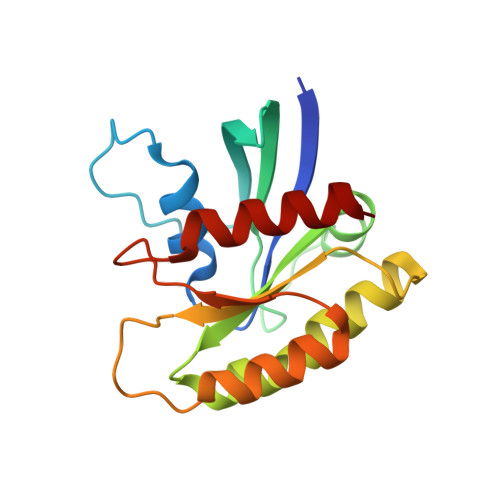
    GTP-bound Ras adopts two interconverting conformations, "inactive" state 1 and "active" state 2. However, the tertiary structure of wild-type (WT) state 1 remains unsolved. Here we solve the state 1 crystal structures of H-Ras WT together with its oncogenic G12V and Q61L mutants. They assume open structures characterized by impaired interactions of both Thr-35 in switch I and Gly-60 in switch II with the γ-phosphate of GTP and possess two surface pockets of mutually different shapes unseen in state 2, a potential target for selective inhibitor development. Furthermore, they provide a structural basis for the low GTPase activity of state 1.


  • Organizational Affiliation
    • Division of Molecular Biology, Department of Biochemistry and Molecular Biology, Kobe University Graduate School of Medicine, 7-5-1 Kusunoki-cho, Chuo-ku, Kobe 650-0017, Japan.

Macromolecule Content 

  • Total Structure Weight: 19.82 kDa 
  • Atom Count: 1,443 
  • Modeled Residue Count: 166 
  • Deposited Residue Count: 171 
  • Unique protein chains: 1

Macromolecules

Find similar proteins by:|  3D Structure
Entity ID: 1
MoleculeChains  Sequence LengthOrganismDetailsImage
GTPase HRas171Homo sapiensMutation(s): 1 
Gene Names: HRASHRAS1
EC: 3.6.5.2
UniProt & NIH Common Fund Data Resources
Find proteins for P01112 (Homo sapiens)
Explore P01112 
Go to UniProtKB:  P01112
PHAROS:  P01112
GTEx:  ENSG00000174775 
Entity Groups
Sequence Clusters30% Identity50% Identity70% Identity90% Identity95% Identity100% Identity
UniProt GroupP01112
Sequence Annotations
Expand
Reference Sequence

Small Molecules

Ligands 2 Unique
IDChains Name / Formula / InChI Key2D Diagram3D Interactions
GNP

Query on GNP


Download:Ideal Coordinates CCD File
B [auth A]PHOSPHOAMINOPHOSPHONIC ACID-GUANYLATE ESTER
C10 H17 N6 O13 P3
UQABYHGXWYXDTK-UUOKFMHZSA-N
MG

Query on MG


Download:Ideal Coordinates CCD File
C [auth A]MAGNESIUM ION
Mg
JLVVSXFLKOJNIY-UHFFFAOYSA-N

Experimental Data & Validation

Experimental Data

  • Method: X-RAY DIFFRACTION
  • Resolution: 2.30 Å
  • R-Value Free:  0.237 (Depositor), 0.233 (DCC) 
  • R-Value Work:  0.183 (Depositor), 0.181 (DCC) 
  • R-Value Observed: 0.185 (Depositor) 
Space Group: I 2 2 2
Unit Cell:
Length ( Å )Angle ( ˚ )
a = 34.782α = 90
b = 82.006β = 90
c = 121.251γ = 90
Software Package:
Software NamePurpose
BSSdata collection
REFMACrefinement
MOSFLMdata reduction
SCALAdata scaling

Structure Validation

View Full Validation Report


Ligand Structure Quality Assessment 


Entry History 

Deposition Data

Revision History  (Full details and data files)

  • Version 1.0: 2012-05-16
    Type: Initial release
  • Version 1.1: 2014-03-12
    Changes: Database references
  • Version 1.2: 2023-11-08
    Changes: Data collection, Database references, Derived calculations, Refinement description