4E70 | pdb_00004e70

Crystal Structure Analysis of Coniferyl Alcohol 9-O-Methyltransferase from Linum Nodiflorum in Complex with Coniferyl Alcohol


Experimental Data Snapshot

  • Method: X-RAY DIFFRACTION
  • Resolution: 1.61 Å
  • R-Value Free: 
    0.191 (Depositor), 0.197 (DCC) 
  • R-Value Work: 
    0.170 (Depositor) 
  • R-Value Observed: 
    0.171 (Depositor) 

wwPDB Validation 3D Report Full Report

Validation slider image for 4E70

Ligand Structure Quality Assessment 


This is version 1.2 of the entry. See complete history

Literature

Structural analysis of coniferyl alcohol 9-O-methyltransferase from Linum nodiflorum reveals a novel active-site environment.

Wolters, S.Neeb, M.Berim, A.Schulze Wischeler, J.Petersen, M.Heine, A.

(2013) Acta Crystallogr D Biol Crystallogr 69: 888-900

  • DOI: https://doi.org/10.1107/S0907444913002874
  • Primary Citation Related Structures: 
    4E70, 4EMS, 4EVI

  • PubMed Abstract: 

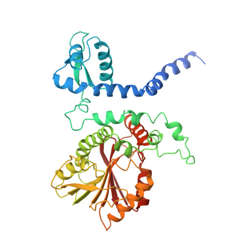
    Coniferyl alcohol 9-O-methyltransferase from Linum nodiflorum (Linaceae) catalyzes the unusual methylation of the side-chain hydroxyl group of coniferyl alcohol. The protein was heterologously expressed in Escherichia coli as a hexahistidine derivative and purified for crystallization. Diffracting crystals were obtained of the pure protein and of its selenomethionine derivative, as well as of complexes with coniferyl alcohol and with S-adenosyl-L-homocysteine together with coniferyl alcohol 9-O-methyl ether (PDB entries 4ems, 4e70 and 4evi, respectively). The X-ray structures show that the phenylpropanoid binding mode differs from other phenylpropanoid O-methyltransferases such as caffeic acid O-methyltransferase. Moreover, the active site lacks the usually conserved and catalytic histidine residue and thus implies a different reaction mode for methylation. Site-directed mutagenesis was carried out to identify critical amino acids. The binding order of coniferyl alcohol and S-adenosyl-L-methionine was investigated by isothermal titration calorimetry experiments.


  • Organizational Affiliation
    • Institut für Pharmazeutische Biologie und Biotechnologie, Philipps-Universität Marburg, Deutschhausstrasse 17A, D-35037 Marburg, Germany.

Macromolecule Content 

  • Total Structure Weight: 87.49 kDa 
  • Atom Count: 6,329 
  • Modeled Residue Count: 715 
  • Deposited Residue Count: 776 
  • Unique protein chains: 1

Macromolecules

Find similar proteins by:|  3D Structure
Entity ID: 1
MoleculeChains  Sequence LengthOrganismDetailsImage
Coniferyl alcohol 9-O-methyltransferase
A, B
388Linum nodiflorumMutation(s): 0 
Gene Names: CA9OMT
UniProt
Find proteins for A6XNE6 (Linum nodiflorum)
Explore A6XNE6 
Go to UniProtKB:  A6XNE6
Entity Groups
Sequence Clusters30% Identity50% Identity70% Identity90% Identity95% Identity100% Identity
UniProt GroupA6XNE6
Sequence Annotations
Expand
Reference Sequence

Experimental Data & Validation

Experimental Data

  • Method: X-RAY DIFFRACTION
  • Resolution: 1.61 Å
  • R-Value Free:  0.191 (Depositor), 0.197 (DCC) 
  • R-Value Work:  0.170 (Depositor) 
  • R-Value Observed: 0.171 (Depositor) 
Space Group: P 21 21 21
Unit Cell:
Length ( Å )Angle ( ˚ )
a = 76.018α = 90
b = 104.451β = 90
c = 109.8γ = 90
Software Package:
Software NamePurpose
SCALEPACKdata scaling
PHENIXrefinement
PDB_EXTRACTdata extraction
MAR345dtbdata collection
HKL-2000data reduction
HKL-2000data scaling
PHASERphasing

Structure Validation

View Full Validation Report


Ligand Structure Quality Assessment 


Entry History 

Deposition Data

Revision History  (Full details and data files)

  • Version 1.0: 2013-05-01
    Type: Initial release
  • Version 1.1: 2013-05-15
    Changes: Database references
  • Version 1.2: 2024-02-28
    Changes: Data collection, Database references, Derived calculations