4E53 | pdb_00004e53

Calmodulin and Nm peptide complex


Experimental Data Snapshot

  • Method: X-RAY DIFFRACTION
  • Resolution: 2.69 Å
  • R-Value Free: 
    0.318 (Depositor), 0.365 (DCC) 
  • R-Value Work: 
    0.276 (Depositor), 0.321 (DCC) 
  • R-Value Observed: 
    0.279 (Depositor) 

wwPDB Validation 3D Report Full Report

Validation slider image for 4E53

This is version 1.2 of the entry. See complete history

Literature

Structural basis for the interaction of unstructured neuron specific substrates neuromodulin and neurogranin with calmodulin

Kumar, V.Chichili, V.P.R.Zhong, L.Tang, X.Velazquez-Campoy, A.Sheu, F.-S.Seetharaman, J.Gerges, N.Z.Sivaraman, J.

(2013) Sci Rep 3: 1392-1392

  • DOI: https://doi.org/10.1038/srep01392
  • Primary Citation Related Structures: 
    4E50, 4E53

  • PubMed Abstract: 

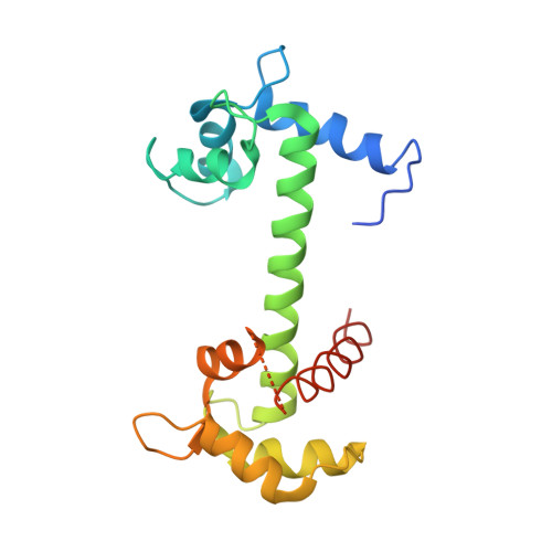
    Neuromodulin (Nm) and neurogranin (Ng) are neuron-specific substrates of protein kinase C (PKC). Their interactions with Calmodulin (CaM) are crucial for learning and memory formation in neurons. Here, we report the structure of IQ peptides (24aa) of Nm/Ng complexed with CaM and their functional studies with full-length proteins. Nm/Ng and their respective IQ peptides are intrinsically unstructured; however, upon binding with CaM, IQ motifs adopt a helical conformation. Ser41 (Ser36) of Nm (Ng) is located in a negatively charged pocket in the apo CaM and, when phosphorylated, it will repel Nm/Ng from CaM. These observations explain the mechanism by which PKC-induced Ser phosphorylation blocks the association of Nm/Ng with CaM and interrupts several learning- and memory-associated functions. Moreover, the present study identified Arg as a key CaM interacting residue from Nm/Ng. This residue is crucial for CaM-mediated function, as evidenced by the inability of the Ng mutant (Arg-to-Ala) to potentiate synaptic transmission in CA1 hippocampal neurons.


  • Organizational Affiliation
    • Department of Biological Sciences, National University of Singapore, Singapore.

Macromolecule Content 

  • Total Structure Weight: 41.49 kDa 
  • Atom Count: 2,715 
  • Modeled Residue Count: 336 
  • Deposited Residue Count: 370 
  • Unique protein chains: 1

Macromolecules

Find similar proteins by:|  3D Structure
Entity ID: 1
MoleculeChains  Sequence LengthOrganismDetailsImage
Calmodulin, Linker, IQ motif of Neuromodulin
A, B
185Mus musculusMutation(s): 0 
UniProt & NIH Common Fund Data Resources
Find proteins for P06837 (Mus musculus)
Explore P06837 
Go to UniProtKB:  P06837
IMPC:  MGI:95639
Find proteins for P0DP26 (Mus musculus)
Explore P0DP26 
Go to UniProtKB:  P0DP26
Entity Groups
Sequence Clusters30% Identity50% Identity70% Identity90% Identity95% Identity100% Identity
UniProt GroupsP0DP26P06837
Sequence Annotations
Expand
Reference Sequence

Experimental Data & Validation

Experimental Data

  • Method: X-RAY DIFFRACTION
  • Resolution: 2.69 Å
  • R-Value Free:  0.318 (Depositor), 0.365 (DCC) 
  • R-Value Work:  0.276 (Depositor), 0.321 (DCC) 
  • R-Value Observed: 0.279 (Depositor) 
Space Group: C 1 2 1
Unit Cell:
Length ( Å )Angle ( ˚ )
a = 79.386α = 90
b = 79.285β = 90.19
c = 136.064γ = 90
Software Package:
Software NamePurpose
HKL-2000data collection
SHELXSphasing
REFMACrefinement
HKL-2000data reduction
HKL-2000data scaling

Structure Validation

View Full Validation Report


Entry History 

Deposition Data

Revision History  (Full details and data files)

  • Version 1.0: 2013-03-20
    Type: Initial release
  • Version 1.1: 2017-06-28
    Changes: Source and taxonomy
  • Version 1.2: 2024-03-20
    Changes: Data collection, Database references