4E3I | pdb_00004e3i

Crystal structure of AmpC beta-lactamase in complex with a designed 3-carboxyl benzyl sulfonamide boronic acid inhibitor


Experimental Data Snapshot

  • Method: X-RAY DIFFRACTION
  • Resolution: 1.60 Å
  • R-Value Free: 
    0.189 (Depositor), 0.186 (DCC) 
  • R-Value Work: 
    0.164 (Depositor), 0.160 (DCC) 
  • R-Value Observed: 
    0.165 (Depositor) 

Starting Model: experimental
View more details

wwPDB Validation 3D Report Full Report

Validation slider image for 4E3I

Ligand Structure Quality Assessment 


This is version 1.3 of the entry. See complete history

Literature

Fragment-guided design of subnanomolar beta-lactamase inhibitors active in vivo.

Eidam, O.Romagnoli, C.Dalmasso, G.Barelier, S.Caselli, E.Bonnet, R.Shoichet, B.K.Prati, F.

(2012) Proc Natl Acad Sci U S A 109: 17448-17453

  • DOI: https://doi.org/10.1073/pnas.1208337109
  • Primary Citation Related Structures: 
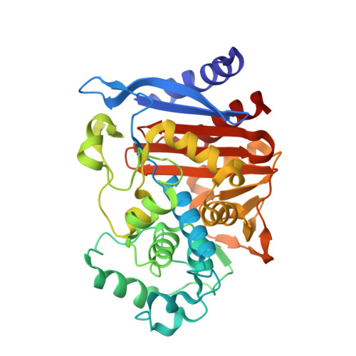
    4E3I, 4E3J, 4E3K, 4E3L, 4E3M, 4E3N, 4E3O

  • PubMed Abstract: 

    Fragment-based design was used to guide derivatization of a lead series of β-lactamase inhibitors that had heretofore resisted optimization for in vivo activity. X-ray structures of fragments overlaid with the lead suggested new, unanticipated functionality and points of attachment. Synthesis of three derivatives improved affinity over 20-fold and improved efficacy in cell culture. Crystal structures were consistent with the fragment-based design, enabling further optimization to a K(i) of 50 pM, a 500-fold improvement that required the synthesis of only six derivatives. One of these, compound 5, was tested in mice. Whereas cefotaxime alone failed to cure mice infected with β-lactamase-expressing Escherichia coli, 65% were cleared of infection when treated with a cefotaxime:5 combination. Fragment complexes offer a path around design hurdles, even for advanced molecules; the series described here may provide leads to overcome β-lactamase-based resistance, a key clinical challenge.


  • Organizational Affiliation
    • Department of Pharmaceutical Chemistry, University of California, 1700 Fourth Street, Byers Hall, San Francisco, CA 94158, USA.

Macromolecule Content 

  • Total Structure Weight: 80.2 kDa 
  • Atom Count: 6,336 
  • Modeled Residue Count: 715 
  • Deposited Residue Count: 716 
  • Unique protein chains: 1

Macromolecules

Find similar proteins by:|  3D Structure
Entity ID: 1
MoleculeChains  Sequence LengthOrganismDetailsImage
Beta-lactamase
A, B
358Escherichia coli K-12Mutation(s): 0 
Gene Names: ampAampCb4150JW4111
EC: 3.5.2.6
UniProt
Find proteins for P00811 (Escherichia coli (strain K12))
Explore P00811 
Go to UniProtKB:  P00811
Entity Groups
Sequence Clusters30% Identity50% Identity70% Identity90% Identity95% Identity100% Identity
UniProt GroupP00811
Sequence Annotations
Expand
Reference Sequence

Experimental Data & Validation

Experimental Data

  • Method: X-RAY DIFFRACTION
  • Resolution: 1.60 Å
  • R-Value Free:  0.189 (Depositor), 0.186 (DCC) 
  • R-Value Work:  0.164 (Depositor), 0.160 (DCC) 
  • R-Value Observed: 0.165 (Depositor) 
Space Group: C 1 2 1
Unit Cell:
Length ( Å )Angle ( ˚ )
a = 118.46α = 90
b = 77.14β = 116.67
c = 97.75γ = 90
Software Package:
Software NamePurpose
XSCALEdata scaling
PHENIXrefinement
PDB_EXTRACTdata extraction

Structure Validation

View Full Validation Report


Ligand Structure Quality Assessment 


Entry History 

Deposition Data

Revision History  (Full details and data files)

  • Version 1.0: 2012-09-26
    Type: Initial release
  • Version 1.1: 2012-11-07
    Changes: Database references
  • Version 1.2: 2023-09-13
    Changes: Data collection, Database references, Derived calculations, Refinement description
  • Version 1.3: 2024-11-06
    Changes: Structure summary